Korean J Gastroenterol.
2014 Dec;64(6):375-379. 10.4166/kjg.2014.64.6.375.
A Case of Low-grade Fibromyxoid Sarcoma of the Colon
- Affiliations
-
- 1Department of Internal Medicine, Ilsan Paik Hospital, Inje University College of Medicine, Goyang, Korea.
- 2Department of Internal Medicine, Asan Medical Center, University of Ulsan College of Medicine, Seoul, Korea. medi01@naver.com
- 3Department of General Surgery, Ilsan Paik Hospital, Inje University College of Medicine, Goyang, Korea.
- 4Department of Pathology, Ilsan Paik Hospital, Inje University College of Medicine, Goyang, Korea.
- KMID: 2160671
- DOI: http://doi.org/10.4166/kjg.2014.64.6.375
Abstract
- Low-grade fibromyxoid sarcoma is a slowly growing soft tissue neoplasm that shows benign histologic features but may have clinical course of malignant disease. It has been reported to occur in the thigh, inguinal area, axilla, shoulder, neck, perineum or buttock. However, there have been few cases of abdominal organ involvement. A 21-year-old woman presented with a large palpable abdominal mass. A 7x4 cm sized round soft tissue tumor at right upper quadrant area was identified by abdominopelvic CT scan. Percutaneous ultrasound-guided biopsy revealed features of spindle cell tumor. On exploration, the tumor originated from transvers colon and was attached to gastrocolic ligament, transverse mesocolon and stomach. The tumor could be dissected with transverse colectomy and partial gastrectomy. The excised tumor, measuring 7x5x5 cm, was well demarcated and appeared as an ovoid mass with firm and myxoid cut surface. She was diagnosed with low-grade fibromyxoid sarcoma arising from transverse colon, and is currently being followed-up without recurrence or metastasis.
Keyword
MeSH Terms
Figure
-
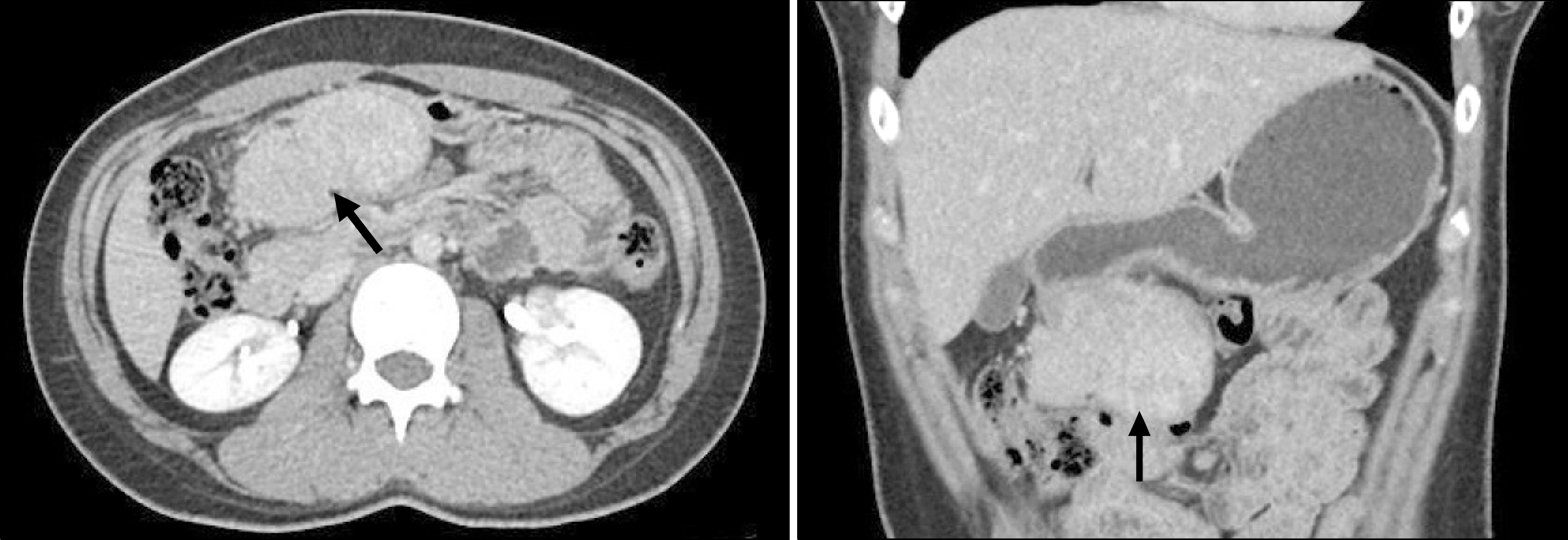
Fig. 1. Abdominal computed tomography shows a 7×4 cm sized homogeneous enhancing mass (arrows) between stomach and transvers colon.
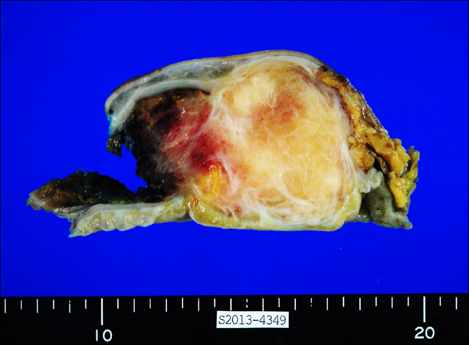
Fig. 2. Gross pathologic image shows ovoid well demarcated lesion and bisecting surface demonstrates a mixture of yellow colored, mucoid and fibrous tissue.

Fig. 3. (A) Transition from fibrous (arrow) to myxoid (asterisk) tumor areas are noted (H&E, ×100). (B) Portion of fibrous tumor reveals arcades of slender blood vessels (H&E, ×100).
Reference
-
References
1. Evans HL. Low-grade fibromyxoid sarcoma. A report of two meta-stasizing neoplasms having a deceptively benign appearance. Am J Clin Pathol. 1987; 88:615–619.
Article2. Evans HL. Low-grade fibromyxoid sarcoma. A report of 12 cases. Am J Surg Pathol. 1993; 17:595–600.3. Goodlad JR, Mentzel T, Fletcher CD. Low grade fibromyxoid sarcoma: clinicopathological analysis of eleven new cases in support of a distinct entity. Histopathology. 1995; 26:229–237.
Article4. Devaney DM, Dervan P, O'Neill S, Carney D, Leader M. Low-grade fibromyxoid sarcoma. Histopathology. 1990; 17:463–465.
Article5. Tang Z, Zhou ZH, Lv CT, et al. Low-grade fibromyxoid sarcoma: clinical study and case report. J Oral Maxillofac Surg. 2010; 68:873–884.
Article6. Kim YJ, Kim KH, Kim KH, et al. Low-grade fibromyxoid sarcoma from the splenic flexure of colon. Korean J Clin Oncol. 2009; 5:69–73.
Article7. Park IJ, Kim HC, Yu CS, Kim JS, Jang SJ, Kim JC. Low-grade fibromyxoid sarcoma of the colon. Dig Liver Dis. 2007; 39:274–277.
Article8. Folpe AL, Lane KL, Paull G, Weiss SW. Low-grade fibromyxoid sarcoma and hyalinizing spindle cell tumor with giant rosettes: a clinicopathologic study of 73 cases supporting their identity and assessing the impact of high-grade areas. Am J Surg Pathol. 2000; 24:1353–1360.9. Zamecnik M, Michal M. Low-grade fibromyxoid sarcoma: a report of eight cases with histologic, immunohistochemical, and ultrastructural study. Ann Diagn Pathol. 2000; 4:207–217.10. Takanami I, Takeuchi K, Naruke M. Low-grade fibromyxoid sarcoma arising in the mediastinum. J Thorac Cardiovasc Surg. 1999; 118:970–971.
Article11. Dvornik G, Barbareschi M, Gallotta P, Dalla Palma P. Low grade fibromyxoid sarcoma. Histopathology. 1997; 30:274–276.
Article12. Shidham VB, Ayala GE, Lahaniatis JE, Garcia FU. Low-grade fibromyxoid sarcoma: clinicopathologic case report with review of the literature. Am J Clin Oncol. 1999; 22:150–155.13. Fukunaga M, Ushigome S, Fukunaga N. Low-grade fibromyxoid sarcoma. Virchows Arch. 1996; 429:301–303.
Article14. Oda Y, Takahira T, Kawaguchi K, et al. Low-grade fibromyxoid sarcoma versus low-grade myxofibrosarcoma in the extremities and trunk. A comparison of clinicopathological and immunohistochemical features. Histopathology. 2004; 45:29–38.
Article15. Fujii S, Kawawa Y, Horiguchi S, Kamata N, Kinoshita T, Ogawa T. Low-grade fibromyxoid sarcoma of the small bowel mesentery: computed tomography and magnetic resonance imaging findings. Radiat Med. 2008; 26:244–247.
Article16. Angervall L, Kindblom LG, Merck C. Myxofibrosarcoma. A study of 30 cases. Acta Pathol Microbiol Scand A. 1977; 85A:127–140.17. Weiss SW, Enzinger FM. Myxoid variant of malignant fibrous histiocytoma. Cancer. 1977; 39:1672–1685.
Article18. Evans HL. Low-grade fibromyxoid sarcoma: a clinicopathologic study of 33 cases with long-term follow-up. Am J Surg Pathol. 2011; 35:1450–1462.
- Full Text Links
-
- Actions
-
Cited
- CITED
-
- Close
- Share
-
- Similar articles
-
- Low Grade Fibromyxoid Sarcoma in Thigh
- Low-Grade Fibromyxoid Sarcoma Arising in Posterior Nasal Cavity: Case Report and Review of the Literature
- Low Grade Fibromyxoid Sarcoma in Chest Wall: One case report
- Primary Paravertebral Low-Grade Fibromyxoid Sarcoma
- A Case of Low-Grade Fibromyxoid Sarcoma Arising in the Finger
