Low Grade Fibromyxoid Sarcoma in Thigh
- Affiliations
-
- 1Department of Orthopaedic Surgery, Cheju Halla General Hospital, Jeju, Korea. dominant0526@naver.com
- 2Department of Pathology, Cheju Halla General Hospital, Jeju, Korea.
- KMID: 999408
- DOI: http://doi.org/10.4055/cios.2009.1.4.240
Abstract
- A low grade fibromyxoid sarcoma is a rare soft tissue tumor that has a tendency to develop in the deep soft tissue of young adults and the potential for local recurrence or distant metastasis. There have been several case reports and sporadic reports in the literature. However, only 1 case has been reported in Korea but without a follow-up result. We describe a 54-year-old female patient with a low-grade fibromyxoid sarcoma of the thigh that had been growing slowly for 34 years. A marginal resection of this tumor was performed. Currently, the patient is doing well without evidence of local recurrence or distant metastasis at 5 years after surgery.
Keyword
MeSH Terms
Figure
-
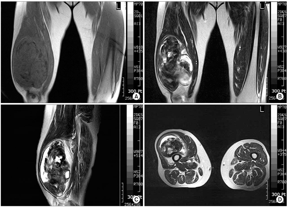
Fig. 1 On MRI, the tumor is a 17 × 11 × 9 cm sized mass located under the quadriceps muscle. The tumor matrix is partially calcified and relatively well defined one having irregular low signal intensity in the T1 weighted image (A) and heterogeneous low signal and high signal in the T2 weighted image (B-D).
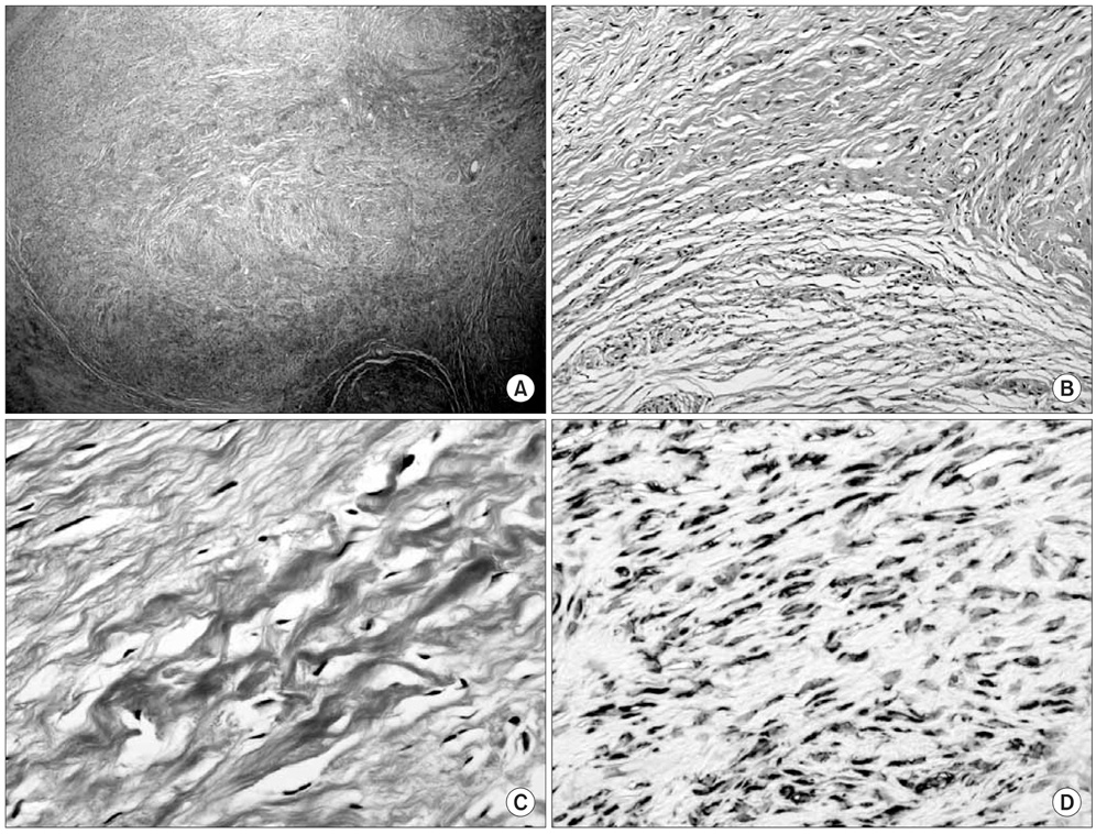
Fig. 2 Optical microscopy examination demonstrates a mass with sharp demarcation, nodular growth pattern and intervening hypocellular collagenous stroma (A: Hematoxylin and eosin stain, ×100). The tumor shows a biphasic pattern with fibrous and myxoid areas, minimal nuclear pleomorphism, low to moderate cellularity, and a swirling, whorled growth (B: Hematoxylin and eosin stain, ×200). The background matrix ranges from fibromyxoid to densely fibrous (C: Hematoxylin and eosin stain, ×400). Immunohistochemically, the tumor cells are diffusely and strongly positive for vimentin (D: Immunohistochemical stain for vimentin, ×1400).
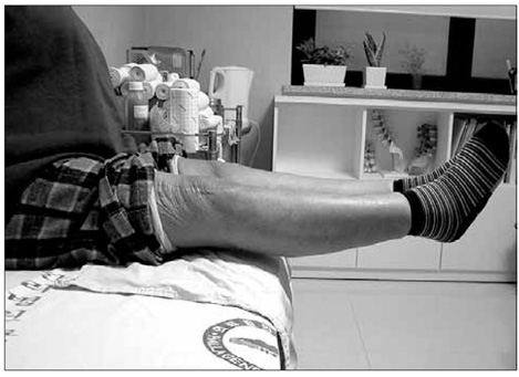
Fig. 3 The patient is still alive with no evidence of local recurrence and distant metastasis five years after excision. The range of motion of the knee joint is full and the strength of the quadriceps muscle is normal.
Reference
-
1. Devaney DM, Dervan P, O'Neill S, Carney D, Leader M. Low-grade fibromyxoid sarcoma. Histopathology. 1990. 17(5):463–465.
Article2. Dvornik G, Barbareschi M, Gallotta P, Dalla Palma P. Low grade fibromyxoid sarcoma. Histopathology. 1997. 30(3):274–276.3. Evans HL. Low-grade fibromyxoid sarcoma: a report of two metastasizing neoplasms having a deceptively benign appearance. Am J Clin Pathol. 1987. 88(5):615–619.
Article4. Evans HL. Low-grade fibromyxoid sarcoma: a report of 12 cases. Am J Surg Pathol. 1993. 17(6):595–600.5. Fukunaga M, Ushigome S, Fukunaga N. Low-grade fibromyxoid sarcoma. Virchows Arch. 1996. 429(4-5):301–303.
Article6. Goodlad JR, Mentzel T, Fletcher CD. Low grade fibromyxoid sarcoma: clinicopathological analysis of eleven new cases in support of a distinct entity. Histopathology. 1995. 26(3):229–237.
Article7. Lee SS, Song C, Sun DH, Moon MS. Low grade fibromyxoid sarcoma in shoulder: one case report. J Korean Bone Joint Tumor Soc. 2004. 10(2):130–133.
- Full Text Links
-
- Actions
-
Cited
- CITED
-
- Close
- Share
-
- Similar articles
-
- Low-Grade Fibromyxoid Sarcoma Arising in Posterior Nasal Cavity: Case Report and Review of the Literature
- A Case of Low-Grade Fibromyxoid Sarcoma with a Typical Clinical Presentation on the Thigh in an Elderly Male
- Primary Paravertebral Low-Grade Fibromyxoid Sarcoma
- Low Grade Fibromyxoid Sarcoma in Chest Wall: One case report
- Cytological Features of Low Grade Fibromyxoid Sarcoma : Report of a Case with a Review of the Literature
