Hyalinizing Spindle Cell Tumor with Giant Rosettes with Pulmonary Metastasis After a Long Hiatus: A Case Report
- Affiliations
-
- 1Department of Clinical Pathology, The Catholic University of Korea, College of Medicine, Seoul, Korea. anhilee@olmh.cuk.ac.kr
- 2Department of Anatomical Pathology, Inha University Hospital, Incheon, Korea.
- KMID: 2157698
- DOI: http://doi.org/10.3346/jkms.2004.19.4.619
Abstract
- "Hyalinizing spindle cell tumor with giant rosettes" (HSCTGR) is a recently described tumor, which is regarded as an unusual variant of low-grade fibromyxoid sarcoma. Proof of a metastatic potential was lacking. The patient in the report was a 35-yr-old woman who showed multiple bilateral pulmonary nodules with massive pleural effusion in the right side. She had a history of a mass excision in the right thigh 11 yrs ago at another hospital, which was reported as a "leiomyoma". Two years before this presentation, the patient received a routine chest radiograph which demonstrated bilateral multiple pulmonary nodules. A lobectomy of the left upper lung was performed. The histological findings revealed a well-circumscribed nodule that was characterized by a spindle-shaped fibrous to hyalinized stroma with criss-crossing short fascicles and giant collagen rosettes surrounded by a rim of spindle-shaped cells. Electron microscopy confirmed the fibroblastic nature of the tumor. This case, in addition to at least two other cases reported in the literature, demonstrates that the HSCTGR is a malignant neoplasm with the capacity to metastasize after a long hiatus.
Keyword
MeSH Terms
Figure
-
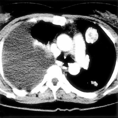
Fig. 1 Computerized tomography of the chest reveals bilateral multiple pulmonary nodules with right massive pleurisy causing collpase of the right lung.

Fig. 2 A fibrous area composed of bland-looking spindle-shaped cells with a fascicular and vague storiform growth pattern. Several rosette-like structures are separated by spindle areas (H&E, ×40).
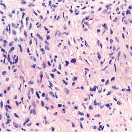
Fig. 3 The tumor cells in the more cellular areas shows irregular wavy nuclei with a moderate degree of nuclear atypia (H&E, ×200).
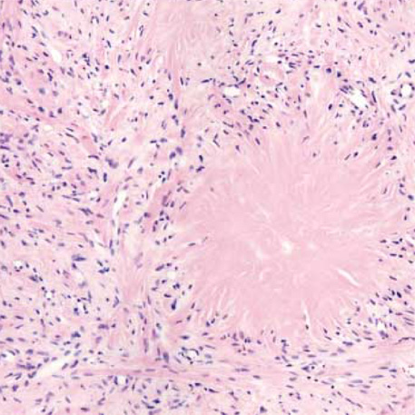
Fig. 4 The central eosinophilic area of the rosettes is composed of collagen fibers (H&E, ×100).

Fig. 5 The thigh tumor shows a moderate cellularity with spindled nuclei in abundant collagenous stroma (H&E, ×200).

Fig. 6 Electron microscopic findings show tumor cells with spindle nuclei containing coarse chromatin, abundant cytoplasm, and dilated rough endoplasmic reticula (×8,000).
Reference
-
1. Lane KL, Shannon RJ, Weiss SW. Hyalinizing spindle cell tumor with giant rosettes. A distinctive tumor closely resembling low-grade fibromyxoid sarcoma. Am J Surg Pathol. 1997. 21:1481–1488.2. Evans HL. Low grade fibromyxoid sarcoma. A report of 12 cases. Am J Surg Pathol. 1993. 17:595–600.3. Woodruff JM, Antonescu CR, Erlandson RA, Boland PJ. Low grade fibrosarcoma with palisaded granuloma-like bodies (giant rosettes). Report of a case that metastasized. Am J Surg Pathol. 1999. 23:1423–1428.4. O'Sullivan MJ, Sirgi KE, Dehner LP. Low-grade fibrosarcoma (hyalinizing spindle cell tumor with giant rosettes) with pulmonary metastases at presentation: case report and review of the literature. Int J Surg Pathol. 2002. 10:211–216.5. Magro G, Fraggetta F, Manusia M, Mingrino A. Hyalinizing spindle cell tumor with giant rosettes. A previously undescribed lesion of the lung. Am J Surg Pathol. 1998. 22:1431–1433.
Article6. Weiss SW, Goldblum JR. Enzinger and Weiss's soft tissue tumors. 2001. 4th ed. St. Louis: Mosby;431–435.7. Folpe AL, Lane KL, Paull G, Weiss SW. Low-grade fibromyxoid sarcoma and hyalinizing spindle cell tumor with giant rosettes. Am J Surg Pathol. 2000. 24:1353–1360.
Article8. Nielsen GP, Selig MK, O'Connell JX, Keel SB, Dickersin GR, Rosenberg AE. Hyalinizing spindle cell tumor with giant rosettes. A report of three cases with ultrastructural analysis. Am J Surg Pathol. 1999. 23:1227–1232.9. Scolyer RA, McCarthy SW, Wills EJ, Palmer AA. Hyalinising spindle cell tumor with giant rosettes: report of a case with unusual features including original histological and ultrastructural observations. Pathology. 2001. 33:101–107.10. Ludvikova M, Michal M, Zamecnik M. Hyalinizing spindle cell tumors with giant rosette-like structures. Pathol Res Pract. 1998. 194:577–581.11. Farinha P, Oliveira P, Soares J. Metastasizing hyalinizing spindle cell tumour with giant rosettes: report of a case with long survival. Histopathology. 2000. 36:92–93.
Article
- Full Text Links
-
- Actions
-
Cited
- CITED
-
- Close
- Share
-
- Similar articles
-
- A Case of Soft Tissue Recurrence after Wide Resection of Giant Cell Tumor in the Distal Femur
- Giant Cell Tumor of the Thoracic Spine Presenting as a Posterior Mediastinal Tumor with Benign Pulmonary Metastases: A Case Report
- A Case of the Giant-Cell Tumor in Coccyx
- Metastasizing Histologically Benign Giant Cell Tumor: A Case Report
- Giant Cell Tumor of the Radius Treated by Massive Resection and fibula Bone graft: One case Report
