J Korean Med Sci.
2004 Aug;19(4):616-618. 10.3346/jkms.2004.19.4.616.
Acquired, Bilateral Nevus of Ota-like Macules (ABNOM) Associated with Ota's Nevus: Case Report
- Affiliations
-
- 1Department of Dermatology, College of Medicine, Kyung Hee University, Seoul, Korea. mhlee@khmc.or.kr
- KMID: 2157697
- DOI: http://doi.org/10.3346/jkms.2004.19.4.616
Abstract
- Ota's nevus is mongolian spot-like macular blue-black or gray-brown patchy pigmentation that most commonly ocurrs in areas innervated by the first and second division of the trigeminal nerve. Acquired, bilateral nevus of Ota-like macules (ABNOM) is located bilaterally on the face, appears later in life, is blue-brown or slate-gray in color. It is not accompanied by macules on the ocular and mucosal membranes. There is also debate as to whether ABNOM is part of the Ota's nevus spectrum. We report an interesting case of ABNOM associated with Ota's nevus. A 36-yr-old Korean women visited our clinic with dark bluish patch on the right cheek and right conjunctiva since birth. She also had mottled brownish macules on both forehead and both lower eyelids that have developed 3 yr ago. Skin biopsy specimens taken from the right cheek and left forehead all showed scattered, bipolar or irregular melanocytes in the dermis. We diagnosed lesion on the right cheek area as Ota's nevus and those on both forehead and both lower eyelids as ABNOM by clinical and histologic findings. This case may support the view that ABNOM is a separate entity from bilateral Ota's nevus.
MeSH Terms
Figure
-

Fig. 1 Dark bluish patch on the right malar area, temple and cheek (Ota's nevus).
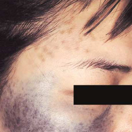
Fig. 2 Mottled brownish macules on both forehead and both lower eyelids (ABNOM lesion).
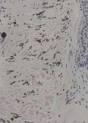
Fig. 3 Skin biopsy from the right malar area (hematoxylin-eosin stain; ×200): Melanocytes, are scattered in the dermis.

Fig. 4 Skin biopsy from the left forehead (hematoxylin-eosin stain; ×200): Melanocytes, are scattered in the dermis.
Reference
-
1. Ota M. Nevus fusco-caeruleus ophthalmo-maxillaris. Jap J Dermatol. 1939. 46:369.2. Hori Y, Kawashima M, Oohara K, Kukita A. Acquired, bilateral nevus of Ota-like macules. J Am Acad Dermatol. 1984. 10:961–964.
Article3. Fitzpatrick TB, Eisen AZ, Wolff K. Dermatology in general medicine. 1987. 3rd ed. New York: McGraw-Hill.4. Mishima Y, Mevorah B. Nevus Ota and nevus Ito in American Negroes. J Invest Dermatol. 1961. 36:133–154.5. Hidano A, Kajima H, Ikeda S, Mizutani H, Miyasato H, Niimura M. Natural history of nevus of Ota. Arch Dermatol. 1967. 95:187–195.
Article6. Hori Y, Takayama O. Circumscribed dermal malanoses; Classification and histologic features. Dermatologic Clinics. 1988. 6:315–326.7. Lowe NJ, Wieder JM, Sawcer D, Burrows P, Chalet M. Nevus of Ota: Treatment with high energy fluences of the Q-switched ruby laser. J Am Acad Dermatol. 1993. 29:997–1001.
Article8. Sun CC, Lu YC, Lee EF, Nakagawa H. Naevus fusco-caeruleus zygomaticus. Br J Dermatol. 1987. 117:545–553.
Article9. Hori Y. Acquired, bilateral nevus of Ota-like macules[reply]. J Am Acad Dermatol. 1985. 12:369.10. Hidano A. Acquired, bilateral nevus of Ota-like macules[letter]. J Am Acad Dermatol. 1985. 12:368–369.
- Full Text Links
-
- Actions
-
Cited
- CITED
-
- Close
- Share
-
- Similar articles
-
- A Case of Acquired Bilateral Nevus of Ota-like Macules Accompanying the Common Blue Nevus
- Tree Cases of Aquired bilateral Nevus of Ota-like Marcules
- Comparison of Characteristics of Acquired Bilateral Nevus of Ota-like Macules and Nevus of Ota According to Therapeutic Outcome
- A Case of Bilateral Nevus of Ota Associated with Bilateral Nevus of Ito
- A Case of Acquired Bilateral Nevus of Ota-like Macules
