Development of a Rat Model of Graded Contusive Spinal Cord Injury Using a Pneumatic Impact Device
- Affiliations
-
- 1Department of Neurosurgery, College of Medicine, Chung-Ang University, Seoul, Korea. tarheelk@hanmail.net
- KMID: 2157688
- DOI: http://doi.org/10.3346/jkms.2004.19.4.574
Abstract
- An animal model of spinal cord trauma is essential for understanding the injury mechanisms, cord regeneration, and to aid the development of new therapeutic modalities. This study focused on the development of a graded experimental contusion model for spinal cord injury (SCI) using a pneumatic impact device made in Korea. A contusive injury was made to the dorsal aspect of the cord. Three trauma groups were defined according to the impact velocity (IV). A control group (n=6), received laminectomy only. Group 1 (n=10), 2 (n=10), and 3 (n=10) had IVs of 1.5 m/sec, 2.0 m/sec, and 3.5 m/sec respectively. Functional assessments were made up to the 14th day after injury. The cord was removed at the 14th post-injury day and prepared for histopathologic examination. Significant behavioral and histopathological abnormalities were found in control and each trauma group. All trauma groups showed severe functional impairment immediately after injury but following different rates of functional recovery (Fig. 5). As the impact velocity and impulse increased, the depth of contusive lesion revealed to be profound the results show that the rat model reproduces spinal cord lesions consistently, has a distinctive value in assessing the effects of impact energy.
MeSH Terms
Figure
-

Fig. 1 Photograph and diagram of pneumatic impact device (CAUH-2). A pneumatic impact controller regulates the flow of high pressure gas to the pneumatic piston. A, Gas; B, Sensor; C, Controller; D, Amplifier; E, Data acquisition board; F, Computer.

Fig. 2 Graph showing the relationship between the driving pressure to the impactor (range, 30-110 psi) and the velocity of the impact tip.

Fig. 3 Graph showing the relationship between the impact velocity and impulse.

Fig. 4 Graph showing the relationship between the impact velocity (A) and impulse (B) in each group. (Value: mean±standard deviation). *p<0.05 vs. group 1, †p<0.05 vs. group 2.
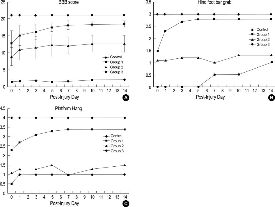
Fig. 5 The time course of functional recovery as measured for the control and experimental groups. (A) The BBB score, (B) Hind foot bar grab, (C) Platform hang.
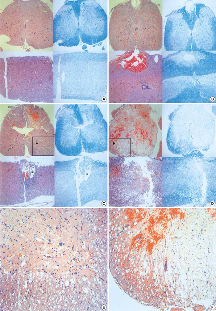
Fig. 6 Representative coronal and sagittal sections of the lesions 14 days after the injury. The dorsal surface is at the top. The left panels are hematoxylin and eosin stain and the right are Luxol fast blue stain (Original magnification, ×40). The Luxol fast blue stain clearly reveals residual myelinated white matter that is restricted to a peripheral rim in the more severe injury groups. (A) control, (B) Group 1, (C) Group 2, (D) Group 3, (E, F) Higher power magnification of the ventral-lateral funiculus (the block demarcates the region of spared white matter, ×100). *: Cystic formation. (E, F) Higher power magnification of the ventral-lateral funiculus (the block demarcates the region of spared white matter, ×100).
Reference
-
1. Andrew RB. Povlishock JT, editor. An overview of spinal cord injury models. Neurotrauma. 1996. New York: McGraw-Hill;1367–1379.2. Seki T, Hida K, Tada M, Koyanagi I, Iwasaki Y. Graded contusion model of the mouse spinal cord using a pneumatic impact device. Neurosurgery. 2002. 50:1075–1081.
Article3. Dixon CE, Clifton GL, Lighthall JW, Yaghmai AA, Hayes RL. A controlled cortical impact model of traumatic brain injury in the rat. J Neurosci Methods. 1991. 39:253–262.4. Lighthall JW. Controlled cortical impact: A new experimental brain injury model. J Neurotrauma. 1988. 5:1–15.
Article5. Lighthall JW, Dixon CE, Anderson TE. Experimental models of brain injury. J Neurotrauma. 1989. 6:83–97.
Article6. Marmarou A, Foda MA, van den Brink W, Campbell J, Kita H, Demetriadou K. A new model of diffuse brain injury in rats. Part I: Pathophysiology and biomechanics. J Neurosurg. 1994. 80:291–300.7. Dixon CE, Lyeth BG, Povlishock JT, Findling RL, Hamm RJ, Marmarou A, Young HF, Hayes RL. A fluid percussion model of experimental brain injury in the rat. J Neurosurg. 1987. 67:110–119.
Article8. Choi SM, Suk JS, Kwon JT, Min BK, Kim YB, Hwang SN, Choi DY, Kim JH, Lee SM, Earmme YY. Development of upgraded cortical impact model (Part I: Mechanics). J Korean Neurosurg Soc. 2002. 32:29–34.9. Choi SM, Suk JS, Min BK, Hwang SN, Kim YB, Kim JH. Development of upgraded cortical impact model (Part II: Functional outcome). J Korean Neurosurg Soc. 2002. 32:458–462.10. Basso DM, Beattie MS, Bresnahan JC. Graded histological and locomotor outocmes after spinal cord contusion using the NYU weight-drop device versus transection. Exp Neurol. 1996. 139:244–256.11. Basso DM, Beattie MS, Bresnahan JC. A sensitive and reliable locomotor rating scale for open field testing in rats. J Neurotrauma. 1995. 12:1–12.
Article12. Kuhn PL, Wrathall JR. A mouse model of graded contusive spinal cord injury. J Neurotrauma. 1998. 15:125–140.
Article13. Bresnahan JC. An electron-microscopic analysis of axonal alterations following blunt contusion of the spinal cord of the rhesus monkey (Macaca mulatta). J Neurol Sci. 1978. 37:59–82.
Article14. Bresnahan JC, King JS, Martin GF, Yashon D. A neuroanatomical analysis of spinal cord injury in the rhesus monkey (Macaca mulatta). J Neurol Sci. 1976. 28:521–542.
Article15. Jakeman LB, Guan Z, Wei P, Ponnappan R, Dzwonczyk R, Popovich PG, Stokes BT. Traumatic spinal cord injury produced by controlled contusion in mouse. J Neurotrauma. 2000. 17:299–319.
Article16. Ma M, Basso DM, Walters P, Strokes BT, Jakeman LB. Behavioral and histological outcomes following graded spinal cord contusion injury in the C57B1/6 mouse. Exp Neurol. 2001. 169:239–254.17. Noble LJ, Wrathall JR. Spinal cord contusion in the rat: Morphometric analyses of alteration in the spinal cord. Exp Neurol. 1985. 88:135–149.
- Full Text Links
-
- Actions
-
Cited
- CITED
-
- Close
- Share
-
- Similar articles
-
- Graded Spinal Cord Contusive Rat Model by a Weight-drop Device
- The Significance of Evoked Potentials according to the Injury Severity of Spinal Cord Contusive Rat Model
- The Effect of the Swimming Exercise on Functional Recovery after Experimental Contusive Spinal Cord Injury in the Rats
- Olig2-expressing Mesenchymal Stem Cells Enhance Functional Recovery after Contusive Spinal Cord Injury
- The Apoptosis and Expression of p53, Bcl-2 in Graded Contusion Injury of Rat Spinal Cord
