Ann Dermatol.
2015 Dec;27(6):787-788. 10.5021/ad.2015.27.6.787.
Spitz Nevus on the Perianal Area of a Child: An Unusual Location
- Affiliations
-
- 1Department of Dermatology, Sanggye Paik Hospital, Inje University College of Medicine, Seoul, Korea. etihwevol@naver.com
- KMID: 2157468
- DOI: http://doi.org/10.5021/ad.2015.27.6.787
Abstract
- No abstract available.
Figure
-

Fig. 1 Spitz nevus on the perianal area: an erythematous pedunculated nodule with tiny black pigmentation anterior to the anal orifice.
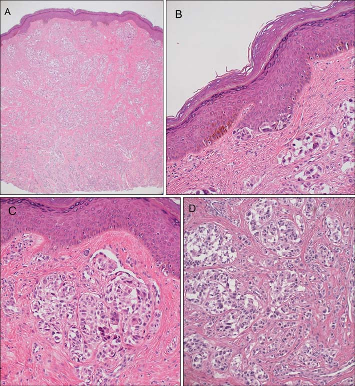
Fig. 2 Histological examination showed (A) numerous nests of cells in the entire dermis (H&E, ×40). On the tip of the rete ridges (B), some proliferation of nevus cells and melanocytes are visible (H&E, ×40). In the superficial dermis (C) and deep dermis (D), the cell nests are composed of epithelioid nevocytes that have a large cell size, abundant cytoplasm, cleft around cells, and prominent nucleoli (H&E, ×200). There was no atypical mitosis or pagetoid spreading.
Reference
-
1. Requena C, Requena L, Kutzner H, Sánchez Yus E. Spitz nevus: a clinicopathological study of 349 cases. Am J Dermatopathol. 2009; 31:107–116.
Article2. Aoyagi S, Sato-Matsumura KC, Akiyama M, Tanimura S, Shibaki H, Shimizu H. Spitz naevus of the glans penis: an unusual location. Acta Derm Venereol. 2004; 84:324–325.
Article3. Filippov SV, Kniaz'kin IV, Anichkov NM, Zeziulin PN, Shinkarenko AV, Bykov NM. Spitz nevus (juvenile nevus) of the penile skin. Arkh Patol. 2002; 64:46–48.4. Cho SB, Kim HS, Jang H. A pedunculated hyalinizing Spitz nevus on the penile shaft. Int J Dermatol. 2009; 48:1134–1136.
Article5. Polat M, Topcuoglu MA, Tahtaci Y, Hapa A, Yilmaz F. Spitz nevus of the genital mucosa. Indian J Dermatol Venereol Leprol. 2009; 75:167–169.
Article
