Ann Dermatol.
2011 Oct;23(Suppl 2):S175-S178. 10.5021/ad.2011.23.S2.S175.
Syringocystadenoma Papilliferum in Co-existence with Tubular Apocrine Adenoma on the Calf
- Affiliations
-
- 1Department of Dermatology, Korea University College of Medicine, Seoul, Korea. drsshong@hanmail.net
- KMID: 2156782
- DOI: http://doi.org/10.5021/ad.2011.23.S2.S175
Abstract
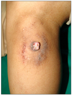
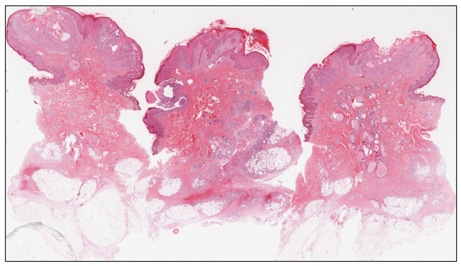
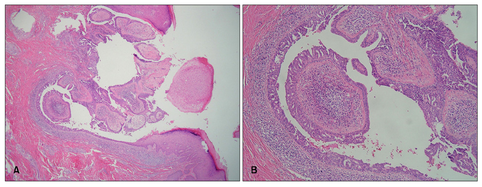
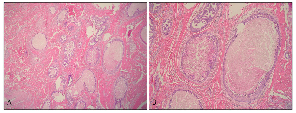
- Syringocystadenoma papilliferum (SCAP) occurs singly or in association with other tumors. Although it is rare, the association of tubular apocrine adenoma (TAA) with SCAP in the background of nevus sebaceous (NS) on the scalp is well documented. However, the co-existence of these two tumors without background of NS has not been reported on the extremities. We report a case of SCAP associated with TAA on the calf without pre-existing NS in an adult.
Figure
Reference
-
1. Mammino JJ, Vidmar DA. Syringocystadenoma papilliferum. Int J Dermatol. 1991. 30:763–766.2. Cribier B, Scrivener Y, Grosshans E. Tumors arising in nevus sebaceus: a study of 596 cases. J Am Acad Dermatol. 2000. 42:263–268.3. Helwig EB, Hackney VC. Syringadenoma papilliferum; lesions with and without naevus sebaceous and basal cell carcinoma. AMA Arch Derm. 1955. 71:361–372.4. Civatte J, Belaïch S, Lauret P. Tubular apocrine adenoma (4 cases) (author's transl). Ann Dermatol Venereol. 1979. 106:665–669.5. Toribio J, Zulaica A, Peteiro C. Tubular apocrine adenoma. J Cutan Pathol. 1987. 14:114–117.
Article6. Ansai S, Watanabe S, Aso K. A case of tubular apocrine adenoma with syringocystadenoma papilliferum. J Cutan Pathol. 1989. 16:230–236.
Article7. Yasuhara M. Typical but rare cases. 1989. Tokyo: Seishindou;22.8. Ahn BK, Park YK, Kim YC. A case of tubular apocrine adenoma with syringocystadenoma papilliferum arising in nevus sebaceus. J Dermatol. 2004. 31:508–510.
Article9. Lee CK, Jang KT, Cho YS. Tubular apocrine adenoma with syringocystadenoma papilliferum arising from the external auditory canal. J Laryngol Otol. 2005. 119:1004–1006.
Article10. Kim MS, Lee JH, Lee WM, Son SJ. A case of tubular apocrine adenoma with syringocystadenoma papilliferum that developed in a nevus sebaceus. Ann Dermatol. 2010. 22:319–322.
Article11. Fujita M, Kobayashi M. Syringocystadenoma papilliferum associated with poroma folliculare. J Dermatol. 1986. 13:480–482.
Article12. Fisher TL. Tubular apocrine adenoma. Arch Dermatol. 1973. 107:137.
Article13. Umbert P, Winkelmann RK. Tubular apocrine adenoma. J Cutan Pathol. 1976. 3:75–87.
Article14. Kazakov DV, Bisceglia M, Calonje E, Hantschke M, Kutzner H, Mentzel T, et al. Tubular adenoma and syringocystadenoma papilliferum: a reappraisal of their relationship. An interobserver study of a series, by a panel of dermatopathologists. Am J Dermatopathol. 2007. 29:256–263.
Article15. Requena L, Kiryu H, Ackerman AB. Requena L, Kiryu H, Ackerman AB, editors. Hamartomas, benign neoplasms. Neoplasms with apocrine differentiation. 1998. Philadelphia: Lippincott-Raven;75–563.16. Hügel H, Requena L. Ductal carcinoma arising from a syringocystadenoma papilliferum in a nevus sebaceus of Jadassohn. Am J Dermatopathol. 2003. 25:490–493.
Article17. Yamane N, Kato N, Yanagi T, Osawa R. Naevus sebaceus on the female breast accompanied with a tubular apocrine adenoma and a syringocystadenoma papilliferum. Br J Dermatol. 2007. 156:1397–1399.
Article
- Full Text Links
- Actions
-
Cited
- CITED
-
- Close
- Share
- Similar articles
-
- Syringocystadenoma Papilliferum of the Back Combined with a Tubular Apocrine Adenoma
- A Case of Tubular Apocrine Adenoma with Syringocystadenoma Papilliferum that Developed in a Nevus Sebaceus
- A Case of Tubular Apocrine Adenoma
- A Cases of Nevus Sebaceus of Jandassohn Associated with Tubular Apocrine Adenoma
- Three Cases Showing Combined Features of Syringocystadenoma Papilliferum andTubular Apocrine Adenoma Superimposed on Nevus Sebaceus


