Whitening Effects of Marine Pseudomonas Extract
- Affiliations
-
- 1Department of Dermatology, Ajou University School of Medicine, Suwon, Korea. hykang@ajou.ac.kr
- 2Department of Dermatology, School of Medicine, Gyeongsang National University, Jinju, Korea.
- 3ShinWon Scientific Co. Ltd, Gunpo, Korea.
- KMID: 2156654
- DOI: http://doi.org/10.5021/ad.2011.23.2.144
Abstract
- BACKGROUND
Bacteria associated with marine invertebrates are a rich source of bioactive metabolites.
OBJECTIVE
The effects of marine bacteria extracts on pigmentation were investigated to find novel whitening agents.
METHODS
The marine bacteria collected near Gangwha Island in Korea were isolated and extracted using organic solvent. The organic extracts were screened and selected using the cell free tyrosinase activity. The whitening effects of the selected extract were further investigated using cultured melanocytes, cultured skin and in vivo zebrafish. The whitening mechanism of the marine extract was also investigated.
RESULTS
The marine bacterial methylene chloride extract reduced the pigmentation of Melan-a cells, human melanocytes, cultured skin and in vivo zebrafish. The decrease in pigmentation was due to the inhibition of tyrosinase activity and the expression of tyrosinase and microphthalmia-associated transcription factor protein. These bacteria were identified as a novel Pseudomonas species.
CONCLUSION
The methylene chloride extract of marine pseudomonas species possesses a whitening effect. Further chemical isolation and characterization of the active compounds from this marine bacterial extract are needed.
Keyword
MeSH Terms
Figure
-
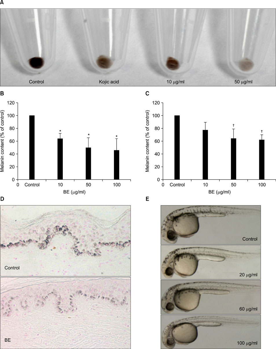
Fig. 1 The bacterial extract (BE) reduced the pigmentation in vitro and in vivo. The extract dose-dependently inhibited the pigmentation in Melan-a cells as shown by the gross appearance of the cell pellets (A) and the decrease in melanin content (B) of the cells. (C) Melanin content of human melanocytes. The values indicate the mean of five independent experiments±SD. *p<0.01, †p<0.05. (D) The bacteria extract (100µg/ml) reduced the basal melanin pigments of cultured human skin as compared to that of the control (Fontana-masson stain, ×200). (E) The effects on the pigmentation of zebrafish observed under the stereomicroscope. Lateral view of the embryos.
Fig. 2 Identification of potential bacteria. The DNA sequencing data revealed that the bacteria belonged to an unidentified Pseudomonas sp.
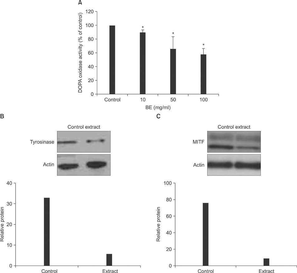
Fig. 3 Effect of the bacterial extract (BE) on the tyrosinase activity and the tyrosinase and MITF expressions. (A) The Melan-a cells were incubated with 10 mM L-DOPA for 90 min and the absorbance was measured at 475 nm. The values indicate the mean of five independent experiments±SD. *p<0.01. The tyrosinase (B) and MITF (C) protein expressions of the Melan-a cells treated with bacterial extract (50µg/ml) were determined by western blotting.
Reference
-
1. Sakuma K, Ogawa M, Sugibayashi K, Yamada K, Yamamoto K. Relationship between tyrosinase inhibitory action and oxidation-reduction potential of cosmetic whitening ingredients and phenol derivatives. Arch Pharm Res. 1999. 22:335–339.
Article2. Jimbow K, Obata H, Pathak MA, Fitzpatrick TB. Mechanism of depigmentation by hydroquinone. J Invest Dermatol. 1974. 62:436–449.
Article3. Faulkner DJ. Marine natural products. Nat Prod Rep. 2002. 19:1–48.
Article4. Proksch P, Edrada RA, Ebel R. Drugs from the seas - current status and microbiological implications. Appl Microbiol Biotechnol. 2002. 59:125–134.
Article5. Lee JS, Lee JY, Choi YM, Jung YS, Kang WH, Kang HY. Skin organ culture model for evaluation of melanin pigmentation. Korean J Dermatol. 2005. 43:450–454.6. Choi TY, Kim JH, Ko DH, Kim CH, Hwang JS, Ahn S, et al. Zebrafish as a new model for phenotype-based screening of melanogenic regulatory compounds. Pigment Cell Res. 2007. 20:120–127.
Article7. Bertolotto C, Buscà R, Abbe P, Bille K, Aberdam E, Ortonne JP, et al. Different cis-acting elements are involved in the regulation of TRP1 and TRP2 promoter activities by cyclic AMP: pivotal role of M boxes (GTCATGTGCT) and of microphthalmia. Mol Cell Biol. 1998. 18:694–702.
Article
- Full Text Links
-
- Actions
-
Cited
- CITED
-
- Close
- Share
-
- Similar articles
-
- Skin Whitening Effects of Sanguisorba officinalis and Stichopus japonicus
- Tooth whitening effects of manicure-type hydrogen peroxide tooth whitening gel
- Tooth Lightness Changes with Listerine Healthy White after Application of Tooth-Coloring-Inducing Foods
- A Clinical Evaluation for the Whitening Effect of the Root of Ma Huang Using Mexameter
- Whitening Effect of Cosmetics Containing Magnesium L-Ascorbyl-2-Phosphate(VC-PMG, Vitamin C Derivatives) Assessed by Colorimeter
