Extraspinal Incidental Findings on Routine MRI of Lumbar Spine: Prevalence and Reporting Rates in 1278 Patients
- Affiliations
-
- 1Department of Radiology, Trakya University Faculty of Medicine, Balkan Campus, Edirne 22000, Turkey. deusedattuncel@yahoo.com
- 2Department of Neurology, Trakya University Faculty of Medicine, Balkan Campus, Edirne 22000, Turkey.
- KMID: 2155561
- DOI: http://doi.org/10.3348/kjr.2015.16.4.866
Abstract
OBJECTIVE
The aim of the present study was to determine the prevalence and reporting rate of incidental findings (IF) in adult outpatients undergoing lumbar magnetic resonance imaging (MRI).
MATERIALS AND METHODS
Re-evaluation of a total of 1278 lumbar MRI images (collected from patients with a mean age of 50.5 years, range 16-91 years) captured between August 2010-August 2011 was done by a neuroradiologist and a musculoskeletal radiologist. IFs were classified according to organ or system (liver, gallbladder, kidney, bladder, uterus, ovary, lymph node, intestine and aorta). The rate of reporting of a range of IF was examined. The outcome of each patient's treatment was evaluated based on review of hospital records and by telephone interviews.
RESULTS
A total of 253 IFs were found in 241 patients (18.8% of 1278). Among these, clinically significant IFs (n = 34) included: 2 renal masses (0.15%), 2 aortic aneurysms (0.15%), 2 cases of hydronephrosis (0.15%), 11 adrenal masses (0.86%), 7 lymphadenopathies (0.55%), 6 cases of endometrial or cervical thickening (0.47%), 1 liver hemangioma (0.08%), 1 pelvic fluid (0.08%) and 2 ovarian dermoid cysts (0.15%). Overall, 28% (71/253) of IFs were included in the clinical reports, while clinically significant findings were reported in 41% (14/34) of cases.
CONCLUSION
Extraspinal IFs are commonly detected during a routine lumbar MRI, and many of these findings are not clinically significant. However, IFs including clinically important findings are occasionally omitted from formal radiological reports.
MeSH Terms
Figure
-
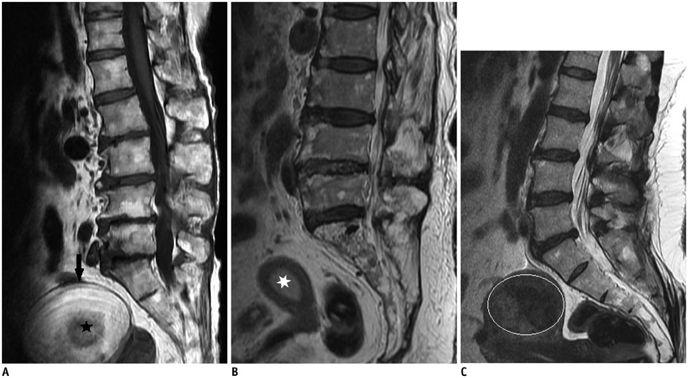
Fig. 1 Sample images from genital system. A. Ovarian dermoid cyst (hyperintense lesion [arrow] on sagittal T1-weighted image) with central dermoid nodule (asterisk), which is verified after surgery. B. Endometrial thickening (asterisk). C. Uterine fibroid (encircled) on T2-weighted sagittal images.

Fig. 2 Pathologies of hepatobiliary, urinary, vascular system and adrenal mass. A. Hepatic hemangioma (arrow). B. Gallbladder stone (arrow). C. Unilateral hydronephrosis (arrow). D. Aortic aneurysm (asterisk). E. Ovoid hyperintense adrenal mass lesion (asterisk) on T2-weighted axial images.
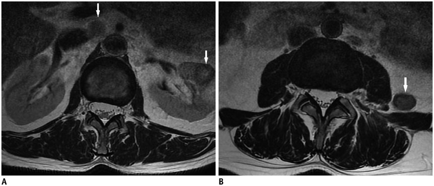
Fig. 3 58-year-old man with three incidental pathologies. A. Renal mass on left kidney and paraaortic enlarged lymph node (arrows). B. Retroperitoneal nodular lesion (arrow) on axial T2-weighted images.
Reference
-
1. Wagner SC, Morrison WB, Carrino JA, Schweitzer ME, Nothnagel H. Picture archiving and communication system: effect on reporting of incidental findings. Radiology. 2002; 225:500–505.2. Fileni A, Magnavita N. A 12-year follow-up study of malpractice claims against radiologists in Italy. Radiol Med. 2006; 111:1009–1022.3. Magnavita N, Magnavita G, Fileni A, Bergamaschi A. Ethical problems in radiology: medical error and disclosure. Radiol Med. 2009; 114:1345–1355.4. Park HJ, Jeon YH, Rho MH, Lee EJ, Park NH, Park SI, et al. Incidental findings of the lumbar spine at MRI during herniated intervertebral disk disease evaluation. AJR Am J Roentgenol. 2011; 196:1151–1155.5. Lee SY, Landis MS, Ross IG, Goela A, Leung AE. Extraspinal findings at lumbar spine CT examinations: prevalence and clinical importance. Radiology. 2012; 263:502–509.6. Hlatky MA, Iribarren C. The dilemma of incidental findings on cardiac computed tomography. J Am Coll Cardiol. 2009; 54:1542–1543.7. Xiong T, Richardson M, Woodroffe R, Halligan S, Morton D, Lilford RJ. Incidental lesions found on CT colonography: their nature and frequency. Br J Radiol. 2005; 78:22–29.8. Konnak JW, Grossman HB. Renal cell carcinoma as an incidental finding. J Urol. 1985; 134:1094–1096.9. Quattrocchi CC, Giona A, Di Martino AC, Errante Y, Scarciolla L, Mallio CA, et al. Extra-spinal incidental findings at lumbar spine MRI in the general population: a large cohort study. Insights Imaging. 2013; 4:301–308.10. Kamath S, Jain N, Goyal N, Mansour R, Mukherjee K. Incidental findings on MRI of the spine. Clin Radiol. 2009; 64:353–361.11. Baysal T, Soylu A. Percutaneous treatment of simple renal cysts with n-butyl cyanoacrylate and iodized oil. Diagn Interv Radiol (Ank). 2009; 15:148–152.12. Pickhardt PJ, Choi JR, Hwang I, Butler JA, Puckett ML, Hildebrandt HA, et al. Computed tomographic virtual colonoscopy to screen for colorectal neoplasia in asymptomatic adults. N Engl J Med. 2003; 349:2191–2200.13. Dahnert W. Radiology review manual. 4th ed. Baltimore: Williams & Wilkins;1999. p. 759.14. Zalis ME, Barish MA, Choi JR, Dachman AH, Fenlon HM, Ferrucci JT, et al. CT colonography reporting and data system: a consensus proposal. Radiology. 2005; 236:3–9.
- Full Text Links
-
- Actions
-
Cited
- CITED
-
- Close
- Share
-
- Similar articles
-
- Utility of Limited Protocol Magnetic Resonance Imaging Lumbar Spine for Nerve Root Compression in a Developing Country, Is It Accurate and Cost Effective?
- Coexisting Spine Lesions on Whole Spine T2 Sagittal MRI in Evaluating Spinal Degenerative Disease
- Incorporation of Whole Spine Screening in Magnetic Resonance Imaging Protocols for Low Back Pain: A Valuable Addition
- The Value of Additional Cervicothoracic Spine Sagittal T2-weighted Images Included in Routine Lumbar Spine MR Imaging
- Cervical Spondylolysis: Report of Two Cases
