Ann Rehabil Med.
2016 Feb;40(1):126-134. 10.5535/arm.2016.40.1.126.
Comparison of Diffusion Tensor Tractography and Motor Evoked Potentials for the Estimation of Clinical Status in Subacute Stroke
- Affiliations
-
- 1Department of Physical and Rehabilitation Medicine, Kangbuk Samsung Hospital, Sungkyunkwan University School of Medicine, Seoul, Korea. kint99@gmail.com
- KMID: 2155175
- DOI: http://doi.org/10.5535/arm.2016.40.1.126
Abstract
OBJECTIVE
To compare diffusion tensor tractography (DTT) and motor evoked potentials (MEPs) for estimation of clinical status in patients in the subacute stage of stroke.
METHODS
Patients with hemiplegia due to stroke who were evaluated using both DTT and MEPs between May 2012 and April 2015 were recruited. Clinical assessments investigated upper extremity motor and functional status. Motor status was evaluated using Medical Research Council grading and the Fugl-Meyer Assessment of upper limb and hand (FMA-U and FMA-H). Functional status was measured using the Modified Barthel Index (MBI). Patients were classified into subgroups according to DTT findings, MEP presence, fractional anisotropy (FA) value, FA ratio (rFA), and central motor conduction time (CMCT). Correlations of clinical assessments with DTT parameters and MEPs were estimated.
RESULTS
Fifty-five patients with hemiplegia were recruited. In motor assessments (FMA-U), MEPs had the highest sensitivity and negative predictive value (NPV) as well as the second highest specificity and positive predictive value (PPV). CMCT showed the highest specificity and PPV. Regarding functional status (MBI), FA showed the highest sensitivity and NPV, whereas CMCT had the highest specificity and PPV. Correlation analysis showed that the resting motor threshold (RMT) ratio was strongly associated with motor status of the upper limb, and MEP parameters were not associated with MBI.
CONCLUSION
DTT and MEPs could be suitable complementary modalities for analyzing the motor and functional status of patients in the subacute stage of stroke. The RMT ratio was strongly correlated with motor status.
Keyword
MeSH Terms
Figure
-
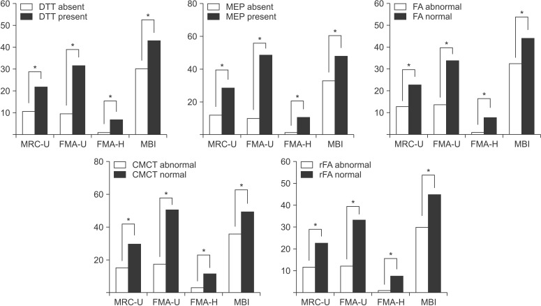
Fig. 1 Comparisons of clinical assessments between subgroups. All clinical assessments showed significant differences between them. MRC-U, Medical Research Council sum of upper limb; FMA-U, Fugl-Meyer Assessment of upper limb; FMA-H, Fugl-Meyer Assessment of hand; DTT, diffusion tensor tractography; FA, fractional anisotropy; rFA, fractional anisotropy ratio; MEP, motor evoked potential; CMCT, central motor conduction time. *p<0.05 by independent t-test.
Reference
-
1. Duncan PW, Goldstein LB, Matchar D, Divine GW, Feussner J. Measurement of motor recovery after stroke. Outcome assessment and sample size requirements. Stroke. 1992; 23:1084–1089. PMID: 1636182.
Article3. York DH. Review of descending motor pathways involved with transcranial stimulation. Neurosurgery. 1987; 20:70–73. PMID: 3543726.
Article4. Thomalla G, Glauche V, Koch MA, Beaulieu C, Weiller C, Rother J. Diffusion tensor imaging detects early Wallerian degeneration of the pyramidal tract after ischemic stroke. Neuroimage. 2004; 22:1767–1774. PMID: 15275932.
Article5. Basser PJ, Pierpaoli C. Microstructural and physiological features of tissues elucidated by quantitativediffusion-tensor MRI. J Magn Reson B. 1996; 111:209–219. PMID: 8661285.
Article6. Lin CC, Tsai MY, Lo YC, Liu YJ, Tsai PP, Wu CY, et al. Reproducibility of corticospinal diffusion tensor tractography in normal subjects and hemiparetic stroke patients. Eur J Radiol. 2013; 82:e610–e616. PMID: 23906441.
Article7. Maraka S, Jiang Q, Jafari-Khouzani K, Li L, Malik S, Hamidian H, et al. Degree of corticospinal tract damage correlates with motor function after stroke. Ann Clin Transl Neurol. 2014; 1:891–899. PMID: 25540803.
Article8. Liu X, Tian W, Qiu X, Li J, Thomson S, Li L, et al. Correlation analysis of quantitative diffusion parameters in ipsilateral cerebral peduncle during Wallerian degeneration with motor function outcome after cerebral ischemic stroke. J Neuroimaging. 2012; 22:255–260. PMID: 21699612.
Article9. Hendricks HT, Hageman G, van Limbeek J. Prediction of recovery from upper extremity paralysis after stroke by measuring evoked potentials. Scand J Rehabil Med. 1997; 29:155–159. PMID: 9271149.10. Udupa K, Chen R. Central motor conduction time. Handb Clin Neurol. 2013; 116:375–386. PMID: 24112910.
Article11. Jang SH, Ahn SH, Sakong J, Byun WM, Choi BY, Chang CH, et al. Comparison of TMS and DTT for predicting motor outcome in intracerebral hemorrhage. J Neurol Sci. 2010; 290:107–111. PMID: 19914639.
Article12. Kwon YH, Son SM, Lee J, Bai DS, Jang SH. Combined study of transcranial magnetic stimulation and diffusion tensor tractography for prediction of motor outcome in patients with corona radiata infarct. J Rehabil Med. 2011; 43:430–434. PMID: 21403983.
Article13. Choi TW, Jang SG, Yang SN, Pyun SB. Factors affecting the motor evoked potential responsiveness and parameters in patients with supratentorial stroke. Ann Rehabil Med. 2014; 38:19–28. PMID: 24639922.
Article14. Liu X, Li L, Tian W, Zhao C, Dogra V. Quantitative diffusion tensor imaging for evaluation of motor function in patients with brain infarcts. Acta Neurol Scand. 2010; 121:315–319. PMID: 20047565.
Article15. Wang DM, Li J, Liu JR, Hu HY. Diffusion tensor imaging predicts long-term motor functional outcome in patients with acute supratentorial intracranial hemorrhage. Cerebrovasc Dis. 2012; 34:199–205. PMID: 23006600.
Article16. Hesse S, Herrmann C, Bardeleben A, Holzgraefe M, Werner C, Wingendorf I, et al. A new orthosis for subluxed, flaccid shoulder after stroke facilitates gait symmetry: a preliminary study. J Rehabil Med. 2013; 45:623–629. PMID: 23804315.
Article17. Fugl-Meyer AR, Jaasko L, Leyman I, Olsson S, Steglind S. The post-stroke hemiplegic patient. 1: a method for evaluation of physical performance. Scand J Rehabil Med. 1975; 7:13–31. PMID: 1135616.18. Sanford J, Moreland J, Swanson LR, Stratford PW, Gowland C. Reliability of the Fugl-Meyer assessment for testing motor performance in patients following stroke. Phys Ther. 1993; 73:447–454. PMID: 8316578.
Article19. Gladstone DJ, Danells CJ, Black SE. The Fugl-Meyer assessment of motor recovery after stroke: a critical review of its measurement properties. Neurorehabil Neural Repair. 2002; 16:232–240. PMID: 12234086.
Article20. Subramanian SK, Yamanaka J, Chilingaryan G, Levin MF. Validity of movement pattern kinematics as measures of arm motor impairment poststroke. Stroke. 2010; 41:2303–2308. PMID: 20814001.
Article21. Duncan PW, Goldstein LB, Horner RD, Landsman PB, Samsa GP, Matchar DB. Similar motor recovery of upper and lower extremities after stroke. Stroke. 1994; 25:1181–1188. PMID: 8202977.
Article22. Broadley SA, Jorgensen L, Cheek A, Salonikis S, Taylor J, Thompson PD, et al. Early investigation and treatment of obstructive sleep apnoea after acute stroke. J Clin Neurosci. 2007; 14:328–333. PMID: 16790349.
Article23. Kunimatsu A, Aoki S, Masutani Y, Abe O, Hayashi N, Mori H, et al. The optimal trackability threshold of fractional anisotropy for diffusion tensor tractography of the corticospinal tract. Magn Reson Med Sci. 2004; 3:11–17. PMID: 16093615.
Article24. Hendricks HT, Pasman JW, van Limbeek J, Zwarts MJ. Motor evoked potentials in predicting recovery from upper extremity paralysis after acute stroke. Cerebrovasc Dis. 2003; 16:265–271. PMID: 12865615.
Article25. Funaba M, Kanchiku T, Imajo Y, Suzuki H, Yoshida Y, Nishida N, et al. Transcranial magnetic stimulation in the diagnosis of cervical compressive myelopathy: comparison with spinal cord evoked potentials. Spine (Phila Pa 1976). 2015; 40:E161–E167. PMID: 25384053.26. Osei-Lah AD, Turner MR, Andersen PM, Leigh PN, Mills KR. A novel central motor conduction abnormality in D90A-homozygous patients with amyotrophic lateral sclerosis. Muscle Nerve. 2004; 29:790–794. PMID: 15170611.
Article27. Lumsden DE, McClelland V, Ashmore J, Charles-Edwards G, Mills K, Lin JP. Central Motor Conduction Time and diffusion tensor imaging metrics in children with complex motor disorders. Clin Neurophysiol. 2015; 126:140–146. PMID: 24836464.
Article28. Weber M, Eisen AA. Magnetic stimulation of the central and peripheral nervous systems. Muscle Nerve. 2002; 25:160–175. PMID: 11870682.
Article29. Pennisi G, Alagona G, Rapisarda G, Nicoletti F, Costanzo E, Ferri R, et al. Transcranial magnetic stimulation after pure motor stroke. Clin Neurophysiol. 2002; 113:1536–1543. PMID: 12350429.
Article30. Homberg V, Stephan KM, Netz J. Transcranial stimulation of motor cortex in upper motor neurone syndrome: its relation to the motor deficit. Electroencephalogr Clin Neurophysiol. 1991; 81:377–388. PMID: 1718724.31. Stulin ID, Savchenko AY, Smyalovskii VE, Musin RS, Stryuk GV, Priz IL, et al. Use of transcranial magnetic stimulation with measurement of motor evoked potentials in the acute period of hemispheric ischemic stroke. Neurosci Behav Physiol. 2003; 33:425–429. PMID: 12921171.32. Liepert J, Miltner WH, Bauder H, Sommer M, Dettmers C, Taub E, et al. Motor cortex plasticity during constraint-induced movement therapy in stroke patients. Neurosci Lett. 1998; 250:5–8. PMID: 9696052.
Article33. Liepert J, Storch P, Fritsch A, Weiller C. Motor cortex disinhibition in acute stroke. Clin Neurophysiol. 2000; 111:671–676. PMID: 10727918.
Article34. Traversa R, Cicinelli P, Bassi A, Rossini PM, Bernardi G. Mapping of motor cortical reorganization after stroke. A brain stimulation study with focal magnetic pulses. Stroke. 1997; 28:110–117. PMID: 8996498.35. Picht T, Strack V, Schulz J, Zdunczyk A, Frey D, Schmidt S, et al. Assessing the functional status of the motor system in brain tumor patients using transcranial magnetic stimulation. Acta Neurochir (Wien). 2012; 154:2075–2081. PMID: 22948747.
Article36. de Oliveira R, Cacho EW, Borges G. Improvements in the upper limb of hemiparetic patients after reaching movements training. Int J Rehabil Res. 2007; 30:67–70. PMID: 17293723.
Article37. Puig J, Pedraza S, Blasco G, Daunis-I-Estadella J, Prats A, Prados F, et al. Wallerian degeneration in the corticospinal tract evaluated by diffusion tensor imaging correlates with motor deficit 30 days after middle cerebral artery ischemic stroke. AJNR Am J Neuroradiol. 2010; 31:1324–1330. PMID: 20299434.
Article
- Full Text Links
-
- Actions
-
Cited
- CITED
-
- Close
- Share
-
- Similar articles
-
- Hand Function and Diffusion Tensor Imaging according to Response of Evoked Potentials in Post-stroke Hemiplegic Patients
- Diffusion Tensor Imaging: Exploring the Motor Networks and Clinical Applications
- Clinical Usefulness of Diffusion Tensor Image Tractography in Stroke Patients: Report of two cases
- Prediction of Motor Recovery Using Diffusion Tensor Tractography in Supratentorial Stroke Patients With Severe Motor Involvement
- Resting-State Metabolism of Hand Knob Area on ¹â¸F-FDG PET-CT According to Hand Function and Tractography of Corticospinal Tract After Stroke
