Invasive Ductal Carcinoma Arising within a Mammary Hamartoma: Case Report
- Affiliations
-
- 1Department of Radiology, Samsung Medical Center, Sungkyunkwan University School of Medicine, Seoul, Korea. claudel@skku.edu
- KMID: 2151771
- DOI: http://doi.org/10.13104/imri.2015.19.4.237
Abstract
- Breast hamartomas are typically a benign condition and rarely develop into malignant lesions. The coexistence of carcinoma and a breast hamartoma is rare; only 15 cases have been reported in the literature. Here, we report a case of invasive ductal carcinoma associated with hamartoma in a 60-year-old woman. Mammography, ultrasonography and magnetic resonance imaging showed typical features of a breast hamartoma and a suspicious mass with microcalcifications arising within the hamartoma.
MeSH Terms
Figure
-
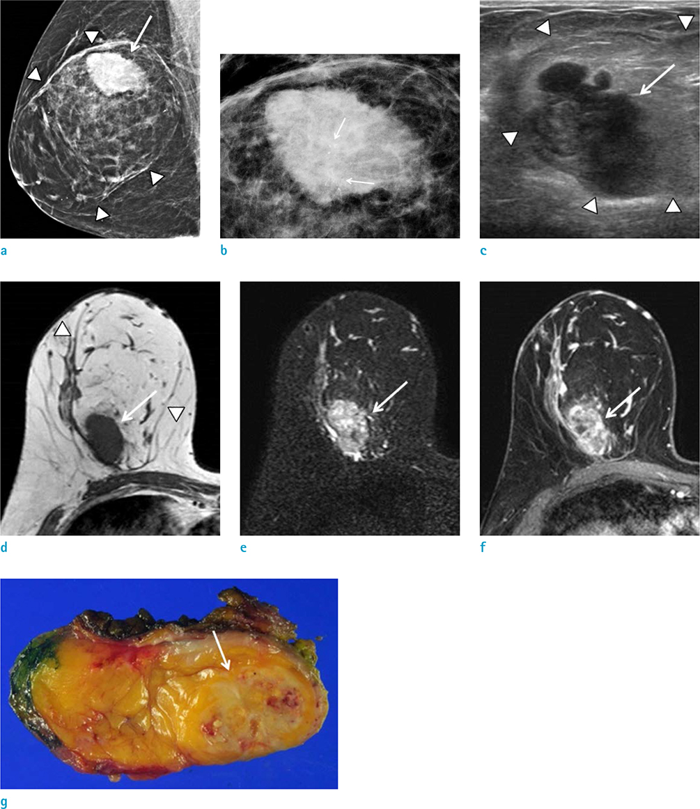
Fig. 1 A 60-year-old woman with a palpable lump in right breast. (a) A right mediolateral oblique mammogram shows a 10 cm, circumscribed, fat-containing mass with a surrounding thin pseudocapsule (arrowheads). Within this fatty mass, in the right upper outer quadrant, a 3.5 cm, irregular hyperdense mass (arrow) is noted. (b) A magnification view of a shows a 3.5 cm, irregular hyperdense mass containing suspicious microcalcifications (arrows) within the fatty mass. (c) Ultrasonography shows a 10 cm, circumscribed oval isoechoic mass in the right upper breast, with a 3.5 cm, irregular hypoechoic mass (arrow) within the large isoechoic mass (arrowheads). (d) Axial precontrast enhanced T1-weighted magnetic resonance (MR) image of the right breast shows a 10 cm, circumscribed, fat-containing mass suggesting breast hamartoma (arrowheads). Within the mass, a 3.5 cm, irregular shaped, isointense mass compared with the muscle is also noted (arrow). (e) T2-weighted MR image of the right breast shows heterogeneous high signal intensity of the 3.5 cm mass (arrow). (f) On axial contrast-enhanced T1-weighted MR image, the mass within the hamartoma shows avid heterogeneous enhancement (arrow). (g) Gross specimen shows a 10.5 × 9 × 5 cm hamartoma with a 3.5 × 3.1 × 2.5 cm mass which appears as a poorly-defined, firm, white-tan, solid mass (arrow).
Reference
-
1. Baron M, Ladonne JM, Gravier A, Picquenot JM, Berry M. Invasive lobular carcinoma in a breast hamartoma. Breast J. 2003; 9:246–248.2. Choi N, Ko ES. Invasive ductal carcinoma in a mammary hamartoma: case report and review of the literature. Korean J Radiol. 2010; 11:687–691.3. Ruiz-Tovar J, Reguero-Callejas ME, Alaez AB, et al. Infiltrating ductal carcinoma and ductal carcinoma in situ associated with mammary hamartoma. Breast J. 2006; 12:368–370.4. Tse GM, Law BK, Ma TK, et al. Hamartoma of the breast: a clinicopathological review. J Clin Pathol. 2002; 55:951–954.5. Kai M, Tada K, Tamura M, et al. Breast cancer associated with mammary hamartoma. Breast Cancer. 2012; 19:183–186.6. Mester J, Simmons RM, Vazquez MF, Rosenblatt R. In situ and infiltrating ductal carcinoma arising in a breast hamartoma. AJR Am J Roentgenol. 2000; 175:64–66.7. Lambert J, Jerjir N, Casselman J, Steyaert L. Invasive lobular carcinoma arising in a hamartoma of the breast: a case report. Clin Breast Cancer. 2015; 15:e63–e66.8. Anani PA, Hessler C. Breast hamartoma with invasive ductal carcinoma. Report of two cases and review of the literature. Pathol Res Pract. 1996; 192:1187–1194.9. Lee EH, Wylie EJ, Bourke AG, Bastiaan De Boer W. Invasive ductal carcinoma arising in a breast hamartoma: two case reports and a review of the literature. Clin Radiol. 2003; 58:80–83.
- Full Text Links
-
- Actions
-
Cited
- CITED
-
- Close
- Share
-
- Similar articles
-
- Invasive Ductal Carcinoma in a Mammary Hamartoma: Case Report and Review of the Literature
- Mammary Hamartoma: A case report
- Synchronous Encapsulated Papillary Carcinoma and Invasive Ductal Carcinoma Arising from Intraductal Papilloma in the Same Breast: A Case Report
- Invasive Ductal Carcinoma Arising in a Recurrent Malignant Phyllodes Tumor: A Case Report
- Co-occurrence of apocrine adenocarcinoma and invasive mammary-type ductal carcinoma in extramammary Paget disease of the axilla
