Invasive Ductal Carcinoma in a Mammary Hamartoma: Case Report and Review of the Literature
- Affiliations
-
- 1Department of Radiology, Konkuk University Medical Center, Konkuk University School of Medicine, Seoul 143-914, Korea. cnm@dreamwiz.com
- 2Department of Radiology, Korea Cancer Center Hospital, Seoul 139-706, Korea.
- KMID: 1119233
- DOI: http://doi.org/10.3348/kjr.2010.11.6.687
Abstract
- Mammary hamartomas are typically a benign condition and rarely develop into malignant lesions. Only 14 cases of carcinomas associated with a hamartoma have been documented in the literature. In this case report, we describe a case of invasive ductal carcinoma within a hamartoma in a 72-year-old woman. Mammography, ultrasonography, and magnetic resonance imaging showed the features of a typical hamartoma with a suspicious mass arising in it. This case illustrates the importance of identification of unusual findings in a typical mammary hamartoma on radiologic examinations.
MeSH Terms
Figure
-
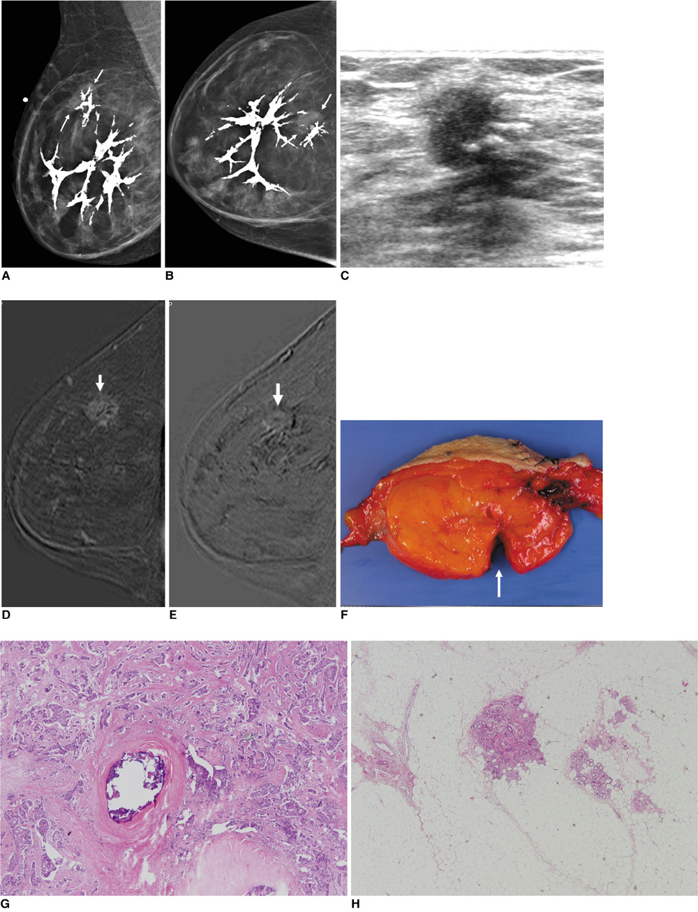
Fig. 1 Invasive ductal carcinoma and mammary hamartoma in 72-year-old woman. A, B. Mediolateral oblique and craniocaudal views of right breast show fat-containing mass including dystrophic calcifications, suggesting hamartoma. There is focal asymmetry (arrows) at 12 o'clock within hamartoma. C. US of right breast at 12 o'clock shows spiculated, nonparallel, hypoechoic mass, which corresponds with focal asymmetry on mammogram (A, B). D, E. Standard subtraction image (D) and reverse subtraction image (E) of dynamic MRI show suspicious mass (arrows) within hamartoma, which was early enhanced (D) and washout (E). F. Gross specimen shows 9-cm smooth circumscribed fatty mass. Suspicious mass was excised from fatty mass (arrow). G, H. Photomicrograph of histopathologic specimen of excised suspicious mass reveals invasive ductal carcinoma and dystrophic calcifications (in H) located within carcinoma.
Reference
-
1. Stavros AT. Breast ultrasound. 2004. Philadelphia: LWW;560–569.2. Mester J, Simmons RM, Vazquez MF, Rosenblatt R. In situ and infiltrating ductal carcinoma arising in a breast hamartoma. AJR Am J Roentgenol. 2000. 175:64–66.3. Mendiola H, Henrik-Nielsen R, Dyreborg U, Blichert-Toft M, Al-Hariri JA. Lobular carcinoma in situ occurring in adenolipoma of the breast. Report of a case. Acta Radiol Diagn (Stockh). 1982. 23:503–505.4. Baron M, Ladonne JM, Gravier A, Picquenot JM, Berry M. Invasive lobular carcinoma in a breast hamartoma. Breast J. 2003. 9:246–248.5. Coyne J, Hobbs FM, Boggis C, Harland R. Lobular carcinoma in a mammary hamartoma. J Clin Pathol. 1992. 45:936–937.6. Kai M, Tada K, Tamura M, Gomi N, Horii R, Akiyama F, et al. Breast cancer associated with mammary hamartoma. Breast Cancer. 2009. [Epub ahead of print].7. Lee EH, Wylie EJ, Bourke AG, Bastiaan De Boer W. Invasive ductal carcinoma arising in a breast hamartoma: two case reports and a review of the literature. Clin Radiol. 2003. 58:80–83.8. Pervatikar SK, Rao R, Dinesh US, Parameswaraiah S. Large mammary hamartoma with focal invasive ductal carcinoma. Indian J Pathol Microbiol. 2009. 52:249–251.9. Ruiz-Tovar J, Reguero-Callejas ME, Aláez AB, Ramiro C, Rojo R, Collado MV, et al. Infiltrating ductal carcinoma and ductal carcinoma in situ associated with mammary hamartoma. Breast J. 2006. 12:368–370.10. Tse GM, Law BK, Pang LM, Cheung HS. Ductal carcinoma in situ arising in mammary hamartoma. J Clin Pathol. 2002. 55:541–542.11. Anani PA, Hessler C. Breast hamartoma with invasive ductal carcinoma. Report of two cases and review of the literature. Pathol Res Pract. 1996. 192:1187–1194.12. Kuroda N, Sugimoto T, Numoto S, Enzan H. Microinvasive lobular carcinoma associated with intraductal spread arising in a mammary hamartoma. J Clin Pathol. 2002. 55:76–77.13. Arrigoni MG, Dockerty MB, Judd ES. The identification and treatment of mammary hamartoma. Surg Gynecol Obstet. 1971. 133:577–582.14. Daya D, Trus T, D'Souza TJ, Minuk T, Yemen B. Hamartoma of the breast, an underrecognized breast lesion. A clinicopathologic and radiographic study of 25 cases. Am J Clin Pathol. 1995. 103:685–689.15. Helvie MA, Adler DD, Rebner M, Oberman HA. Breast hamartomas: variable mammographic appearance. Radiology. 1989. 170:417–421.16. Chao TC, Chao HH, Chen MF. Sonographic features of breast hamartomas. J Ultrasound Med. 2007. 26:447–452.17. Park SY, Oh KK, Kim EK, Son EJ, Chung WH. Sonographic findings of breast hamartoma: emphasis on compressibility. Yonsei Med J. 2003. 44:847–854.
- Full Text Links
-
- Actions
-
Cited
- CITED
-
- Close
- Share
-
- Similar articles
-
- Invasive Ductal Carcinoma Arising within a Mammary Hamartoma: Case Report
- Mammary Hamartoma: A case report
- Invasive Ductal Carcinoma of the Male Breast: A Case Report and Review of the Literature
- Myoid Hamartoma of the Breast: A Case Report
- Co-occurrence of apocrine adenocarcinoma and invasive mammary-type ductal carcinoma in extramammary Paget disease of the axilla
