J Korean Soc Spine Surg.
2015 Dec;22(4):183-185. 10.4184/jkss.2015.22.4.183.
Brain Tumor Mimicking Cervical Spinal Disease: A Case Report
- Affiliations
-
- 1Department of Orthopedic Surgery, School of Medicine, Wonkwang University Hospital, Iksan, Korea.
- 2Department of Orthopedic Surgery, Keimyung University School of Medicine, Daegu, Korea. polo4164@naver.com
- KMID: 2150547
- DOI: http://doi.org/10.4184/jkss.2015.22.4.183
Abstract
- STUDY DESIGN: Case report.
OBJECTIVES
To report a case of cerebellar tumor mimicking cervical spinal disease with neck pain for one year. SUMMARY OF LITERATURE REVIEW: Neck pain is one of the most common symptoms of cervical spinal disease. Neck pain in the cervical spine is usually accompanied by radiculopathic or myelopathic symptoms. Pain aggravated with neck motion is another point of differentiation. However, the differential diagnosis of neck pain is not always easy.
MATERIALS AND METHODS
A 47-year-old woman presented with neck pain, without other symptoms of radiculopathy or myelopathy. The neck pain was not position-dependent and had exacerbated 1 week previously. Cervical magnetic resonance imaging (MRI) revealed a brain tumor in the cerebellum.
RESULTS
The patient underwent surgical craniotomy and tumor resection. The neck pain improved after surgery.
CONCLUSIONS
If neck pain cannot be explained by cervical pathological conditions, the possibility of other causes, including brain pathology, should be considered.
Keyword
MeSH Terms
Figure
-
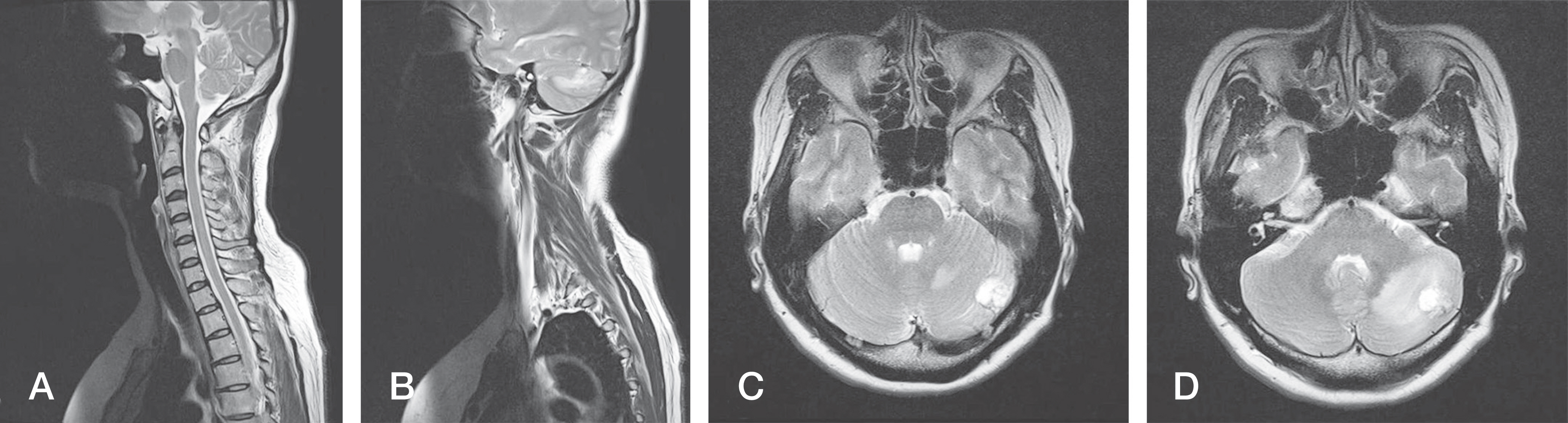
Fig. 1. Cervical spine magnetic resonance imaging (MRI) and brain MRI.(A) T2 sagittal cervical MRI showed no cervical abnormality, (B) T2 sagittal cervical MRI showed high signal intensity in the cerebellum and surrounding edema, (C) T2 axial brain MRI showed a left cerebellar mass with high signal intensity, (D) T2 axial brain MRI showed a left cerebellar mass with high signal intensity and surrounding edema.
Reference
-
1. Kim TK, Shim DM, Oh SK, et al. Diagnostic and Thera-peutic Utility of Ultrasonography-guided Facet Joint Block in Chronic Cervical Spinal Pain. J Korean Orthop US Soc. 2010; 2:54–8.2. Kang CH, Jeon SH, Lee H. Clinical Symptoms of Internal Disc Disruption of the Cervical Spine. J Korean Soc Spine Surg. 2002; 9:48–53.
Article3. Barnsley L, Bogduk N. Medial branch blocks are specific for diagnosis of cervical zygapophyseal joint pain. Reg Anesth. 1993; 18:343–50.4. Therrien AS, Bastian AJ. Cerebellar damage impairs internal predictions for sensory and motor function. Curr Opin Neurobiol. 2015; 33:127–33.
Article5. Stein PJ. A case of cerebellopontine angle meningioma presenting with neck and upper extremity pain. J Manipulative Physiol Ther. 2009; 32:776–80.
Article6. Richard S, Campello C, Taillandier L, et al. Haemangio-blastoma of the central nervous system in von Hippel-Lindau disease. J Intern Med. 1998; 243:547–53.
Article7. Glä sker S, Van Velthoven V. Risk of hemorrhage in heman-gioblastomas of the central nervous system. Neurosurgery. 2005; 57:71–6.
- Full Text Links
-
- Actions
-
Cited
- CITED
-
- Close
- Share
-
- Similar articles
-
- Motion Induced Artifact Mimicking Cervical Dens Fracture on the CT Scan: A Case Report
- Neuromyelitis Optica Spectrum Disorders Mimicking Multiple Brain Tumors
- Charcot Arthropathy of the Lumbosacral Spine Mimicking a Vertebral Tumor after Spinal Cord Injury
- Importance of Differential Diagnosis of a Possible Brain Tumor in Patients with Cervical Radiculopathy
- Primary central nervous system lymphoma in the brainstem and cervical spinal cord: a case report and literature review
