Xanthogranulomatous Pyelonephritis in an Infant
- Affiliations
-
- 1Department of Urology, Ewha Womans University College of Medicine, Seoul, Korea. woochung@ewha.ac.kr
- KMID: 2139785
- DOI: http://doi.org/10.4111/kju.2006.47.12.1367
Abstract
- Xanthogranulomatous pyelonephritis (XGP) is an uncommon, severe, chronic infection of the kidney, which results in the normal renal parenchyma being replaced by characteristic lipid-laden macrophages (foam cells). It can occur at any age, but is extremely rare in children. Herein, the case of a 6-month-3-week-old girl with XGP, pre-operatively diagnosed with a Wilms' tumor, is reported. This is the youngest age of any patient with such a condition reported in the Korean literatures. Excretory urography, abdominal sonogram, computed tomogram and magnetic resonance imaging revealed a right renal round mass, without any calcifications. Her pathological findings after a radical nephrectomy revealed XGP.
Keyword
MeSH Terms
Figure
-
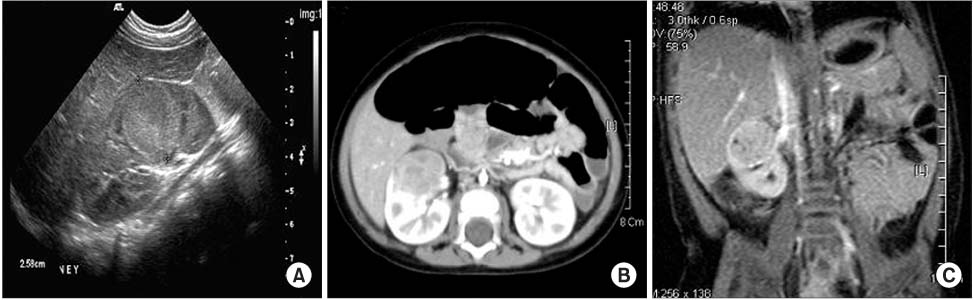
Fig. 1 Radiological findings. (A) Ultrasonography of the abdomen shows a well demarcated heterogeneous echogenic lesion with a thin echogenic rim in the middle portion of the slightly enlarged right kidney. (B) Postcontrast enhancement of abdominal computerized tomography demonstrates a well demarcated, round soft tissue density mass at the upper pole of the right kidney. (C) T1-weighted image, coronal section of a magnetic resonance image shows a heterogeneous, diffuse enhancement and irregularly shaped low signal intensity mass at the anterior aspect of the middle portion of the right kidney.

Fig. 2 Microscopic findings of the specimen reveal extensive pyelonephritis, granulomas, atrophied glomerulus and tubules, and lipid-laden macrophages (foam cells) (black arrow) and plasma cells (white arrow) (H&E, ×200).
Cited by 1 articles
-
Xanthogranulomatous Pyelonephritis in Korean Children
Jong Kil Nam, Sung Woo Park, Sang Don Lee, Moon Kee Chung
Yonsei Med J. 2012;53(6):1159-1164. doi: 10.3349/ymj.2012.53.6.1159.
Reference
-
1. Hammadeh MY, Nicholls G, Calder CJ, Buick RG, Gornall P, Corkery JJ. Xanthogranulomatous pyelonephritis in childhood: pre-operative diagnosis is possible. Br J Urol. 1994. 73:83–86.2. Samuel M, Duffy P, Capps S, Mouriquand P, Williams D, Ransley P. Xanthogranulomatous pyelonephritis in childhood. J Pediatr Surg. 2001. 36:598–601.3. Jeong KS, Kim DS, Cho JH. A cases of xanthogranulomatous pyelonephritis in children. Korean J Urol. 1994. 35:82–85.4. Avnet NL, Roberts TW, Goldberg HR. Temefactive xanthogranulomatous pyelonephritis. Am J Roentgenol Radium Ther Nucl Med. 1963. 90:80–96.5. Choi SH, Lee JH, Cho SR. Xanthogranulomatous pyelonephritis in a child. Korean J Urol. 2005. 46:1231–1234.6. Kural AR, Akaydin A, Oner A, Ozbay G, Solok V, Oruc N, et al. Xanthogranulomatous pyelonephritis in children and adults. Br J Urol. 1987. 59:383–385.7. Kang TW, Jung SI, Jung GW. Clinical studies of xanthogranulomatous pyelonephritis. Korean J Urol. 2001. 42:279–284.8. Dunnick NR, Sandler CM, Amis ES, Newhouse JH. Dunnick NR, Sandler CM, Amis ES, Newhouse JH, editors. Renal inflammatory disease. Textbook of uroradiology. 1997. 2nd ed. Baltimore: Williams & Wilkins;163–189.9. Rasoulpour M, Banco L, Mackay IM, Height DW, Berman MM. Treatment of focal xanthogranulomatous pyelonephritis with antibiotics. J Pediatr. 1984. 105:423–425.10. Sugie S, Tanaka T, Nishikawa A, Yoshimi N, Kato K, Mori H, et al. Fine-needle aspiration cytology of xanthogranulomatous pyelonephritis. Urology. 1991. 37:376–379.
