Asymptomatic Myxoma Originating from the Right Ventricular Outflow Tract
- Affiliations
-
- 1Division of Cardiology, Department of Internal Medicine, College of Medicine, The Catholic University of Korea, Seoul, Korea. jung30134@naver.com
- KMID: 2135418
- DOI: http://doi.org/10.4250/jcu.2013.21.4.186
Abstract
- Asymptomatic right ventricular outflow tract (RVOT) myxoma is quite rare. We report an unusual case of asymptomatic myxoma arising from the RVOT which was successfully surgically removed.
MeSH Terms
Figure
-
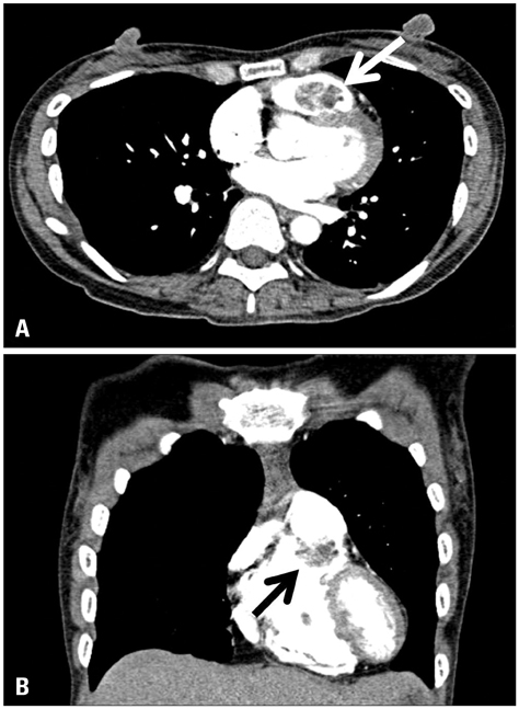
Fig. 1 A: Pulmonary angio computed tomography (CT). CT shows a 2.4 × 1.4 cm sized mass in right ventricular outflow tract (RVOT) (white arrow). B: The mass was attached to the subvalvular infundibulum, extending into the RVOT (black arrow).

Fig. 2 Transthoracic echocardiography. Parasternal short axis view at the level of the aortic valve reveals a 24 × 17 mm sized myxoma (arrow). Ao: aorta, RA: right atrium, RV: right ventricle.
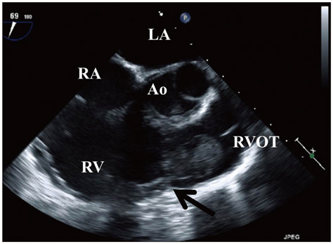
Fig. 3 Transesophageal echocardiography. Transesophageal view shows a mass in the right ventricular outflow tract attached to the infundibulum of the right ventricle (arrow). Ao: aorta, LA: left atrium, RA: right atrium, RV: right ventricle, RVOT: right ventricular outflow tract.
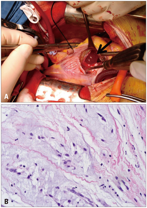
Fig. 4 A: Intraoperative view of the mass (arrow) seen after incising right atrium. B: The microscopic examination shows stellate and globular cells that are arranged in cord-like pattern with abundant myxoid background (H&E, × 400).
Reference
-
1. Gopal AS, Stathopoulos JA, Arora N, Banerjee S, Messineo F. Differential diagnosis of intracavitary tumors obstructing the right ventricular outflow tract. J Am Soc Echocardiogr. 2001; 14:937–940.
Article2. Gajjar TP, Shah GB, Desai NB. Giant ventricular myxoma obstructing right ventricular outflow tract. Rev Bras Cir Cardiovasc. 2011; 26:663–666.3. Kim DC, Song JS, Bae JH, Kim MS, Rho JR. A case of right ventricular myxoma. Korean J Intern Med. 1981; 24:626–638.
Article4. Park JK, Song IS, Lee HK. A case report of giant right ventricular myxoma. Korean J Thorac Cardiovasc Surg. 1983; 16:470–475.5. Song H, Baek WK, Ahn H, Chae H, Kim CW. Surgical excision of intracardiac myxoma: a 15-year experience. Korean J Thorac Cardiovasc Surg. 1992; 25:176–182.6. Min PK, Park BE, Moon JY, Kang SM, Ha JW, Rim SJ, Chang BC, Chung N. Right ventricular myxoma prolapsing into pulmonary artery with significant obstruction. J Korean Soc Echocardiogr. 2003; 11:42–45.
Article7. Paraskevaidis IA, Triantafilou K, Karatzas D, Kremastinos DT. Right ventricular multiple myxomas obstructing right ventricular outflow tract. J Thorac Cardiovasc Surg. 2003; 126:913–914.
Article8. van der Heusen FJ, Stratmann G, Russell IA. Right ventricular myxoma with partial right ventricular outflow tract obstruction. Anesth Analg. 2006; 103:305–306.
Article
- Full Text Links
-
- Actions
-
Cited
- CITED
-
- Close
- Share
-
- Similar articles
-
- Left Ventricular Myxoma Leading to Left Ventricular Outflow Tract Obstruction
- Right Ventricular Myxoma Obstructing Right Ventricular Outflow Tract
- A Rare Case of Unruptured Sinus of Valsalva Aneurysm Obstructing the Right Ventricular Outflow Tract
- Right Ventricular Myxoma Prolapsing into Pulmonary Artery with Significant Obstruction
- Idiopathic Polymorphic Ventricular Tachycardia: a “Benign Disease†with a Touch of Bad Luck?
