Comparison of Damage Degrees After Corneal Epithelial Debridement Using Different Instruments in Rabbit Eyes
- Affiliations
-
- 1Department of Ophthalmology, Wonju Christian Hospital, Yonsei University Wonju College of Medicine, Wonju, Korea. bswhitey@hanmail.net
- 2Yonsei Prime Eye Center, Wonju, Korea.
- KMID: 2122295
- DOI: http://doi.org/10.3341/jkos.2010.51.9.1264
Abstract
- PURPOSE
A corneal epithelial debridement using three different instruments was performed in rabbits, and the rates of corneal epithelium recovery were compared. Additionally, the extent of corneal damage as determined by the scanning electron microscopy was evaluated in each group.
METHODS
Nineteen eyes of ten rabbits were classified into three groups according to the instruments used. The corneal epithelial debridement was performed using three different instruments: a Beaver blade (group A), a Bard-Parker blade No.15 (group B) and a dry cotton-tipped ap plicator (group C). After epithelial debridement, each cornea was observed every 24 hours for three days. After completion of the corneal recovery, each cornea was severed along the limbus and observed under the scanning electron microscope.
RESULTS
The rate of corneal epithelial healing of the group C (dry cotton-tipped applicator) showed no statistical significance from those of the other groups. However, according to the corneal status observed under scanning electron microscope after debridement, cracks in the corneal surface in portions of group A and B were observed in contrast with no creaks in group C.
CONCLUSIONS
Based on these results, corneal epithelial debridement using a cotton-tipped applicator is expected to reduce the occurrence of postoperative corneal complications. Use of a dry cotton-tipped applicator for corneal epithelial debridement in vitreoretinal surgery is suggested.
Keyword
MeSH Terms
Figure
-

Figure 1 Corneal epithelial defect in Group A (Beaver blade group) (Initial, after 24, 48, 72hrs)

Figure 2 Corneal epithelial defect in Group B (Bard-Parker blade No. 15 group) (Initial, after 24, 48, 72hrs)
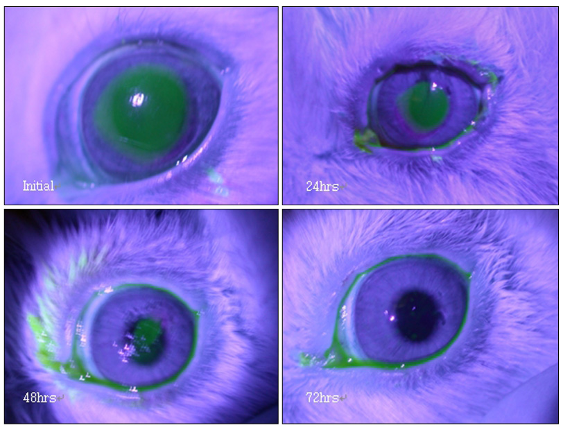
Figure 3 Corneal epithelial defect in Group C (Dry cotton-tipped applicator group) (Initial, after 24, 48, 72hrs)

Figure 4 Corneal epithelial surface after debridement with a dry cotton-tipped applicator: Scanning electronmicroscopy demonstrated normal corneal surface (×350)
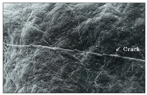
Figure 5 Corneal epithelial surface after debridement with a Beaver blade: Scanning electronmicroscopy demonstrated a crack on the corneal surface (×350)

Figure 6 Corneal epithelial surface after debridement with a Beaver blade: Scanning electronmicroscopy demonstrated a crack on the corneal surface (×350)
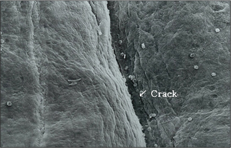
Figure 7 Corneal epithelial surface after debridement with a Bard-Parker blade No.15: Scanning electronmicroscopy demonstrated a crack on the corneal surface (×350)
Reference
-
1. Lee JH. The Retinal Detachment. 1996. 1st ed. Seoul: SNU press;105.2. Chung H. The Eye and the Diabetes Mellitus. 1999. 1st ed. Seoul: SNU press;38–39. 170–171.3. Garcia-Valenzuela E, Abdelsalam A, Eliott D, et al. Reduced need for corneal epithelial debridement during vitreo-retinal surgery using two different viscous surface lubricants. Am J Ophthalmol. 2003. 136:1062–1066.4. Chung H, Tolentino FI, Cajita VN, et al. Reevaluation of corneal complications after closed vitrectomy. Arch Ophthalmol. 1988. 106:916–919.5. Perry HD, Foulks GN, Thoft RA, Tolentino FI. Corneal complications after closed vitrectomy through the pars plana. Arch Ophthalmol. 1978. 96:1401–1403.6. Schachat AP, Oyakawa RT, Michels RG, Rice TA. Complications of vitreous surgery for diabetic retinopathy. II. Postoperative complications. Ophthalmology. 1983. 90:522–530.7. Sánchez-Thorin JC. The cornea in diabetes mellitus. Int Ophthalmol Clin. 1998. 38:19–36.8. Foulks GN, Thoft RA, Perry HD, Tolentino FI. Factors related to corneal epithelial complications after closed vitrectomy in diabetics. Arch Ophthalmol. 1979. 97:1076–1078.9. Kinoshita JH, Fukushi S, Kador P, Merola LO. Aldose reductase in diabetic complications of the eye. Metabolism. 1979. 28:462–469.10. Spencer R, Newsome DA, Schepens CL. Limited superficial debridement to improve corneal clarity during closed vitrectomy. Am J Ophthalmol. 1980. 89:137–138.11. Friberg TR, Ohji M, Scherer JJ, Tano Y. Frequency of epithelial debridement during diabetic vitrectomy. Am J Ophthalmol. 2003. 135:553–554.12. Hong JW, Kim HM, Kang SM. Comparison of Wound Healing and Inflammation Depending on the ablation Methods in Rabbits. J Korean Ophthalmol Soc. 1998. 39:1971–1977.13. Kim EC, Joo CK. Comparison of Clinical Result of Epithelial Removal during Photorefractive Keratectomy with Blade to Brush. J Korean Ophthalmol Soc. 2002. 43:1138–1144.14. KEEDS. The Cornea. 2005. 2nd ed. Seoul: Ilchokak;chap.1-4.15. Kang SM, Song YM, Kim JH. The Factors Related with Epithelial Healing after Vitrectomy for Proliferative Diabetic Retinopathy. J Korean Ophthalmol Soc. 2005. 46:1486–1490.
- Full Text Links
-
- Actions
-
Cited
- CITED
-
- Close
- Share
-
- Similar articles
-
- The Factors Related with Epithelial Healing after Vitrectomy for Proliferative Diabetic Retinopathy
- Comparison of Pain between Ethanol-assisted Mechanical and Conventional Debridement of Corneal Epithelium in PRK
- 20% Alcohol Toxicity on Rabbit Corneal Epithelial Cells: Electron microscopic study
- Inhibition of Corneal Neovascularization by Subconjunctival Injection of alphaVbeta5 Integrin Antibody in Rabbit
- The Effect of RGP Lens and Reverse Geometry Lens on Redistribution of Corneal Epithelial Cell in Rabbit
