J Korean Ophthalmol Soc.
2010 Sep;51(9):1237-1244. 10.3341/jkos.2010.51.9.1237.
Usefulness of Table Parameters of Stratus OCT in Detection of Localized Retinal Nerve Fiber Layer Defects
- Affiliations
-
- 1Department of Ophthalmology, Kang Dong Sacred Heart Hospital, Hallym University College of Medicine, Seoul, Korea. demian7435@gmail.com
- 2Department of Ophthalmology, Seoul National University College of Medicine, Seoul, Korea.
- KMID: 2122291
- DOI: http://doi.org/10.3341/jkos.2010.51.9.1237
Abstract
- PURPOSE
To evaluate the usefulness of table parameters of Stratus optical coherence tomography (OCT) in order to detect localized retinal nerve fiber layer (RNFL) defects.
METHODS
The present study included 86 glaucoma patients with only localized, wedge-shaped RNFL defects, as determined by red-free RNFL photographs. All subjects were tested fast RNFL scans, using of Stratus OCT. The sensitivity of the clock hour parameter and 11 table parameters of RNFL thickness average analysis were compared.
RESULTS
The best parameters in the superior table parameter of the Stratus OCT were Smax, Savg, and Smax/Tavg (sensitivity = 36.7%, 36.7%, 36.7%, respectively). The best parameters in the inferior table parameter of the Stratus OCT were Iavg, Imax, and Imax/Smax (sensitivity = 63.8%, 59.4%, and 50.7%, respectively). However, all were significantly lower than the sensitivity of the clock hour parameter (superior RNFL defect: 60%; inferior RNFL defect: 84.1%).
CONCLUSIONS
The usefulness of the table parameters of the Stratus OCT used to detect localized RNFL defects in glaucoma patients is considered low because of its low sensitivity.
Keyword
Figure
-
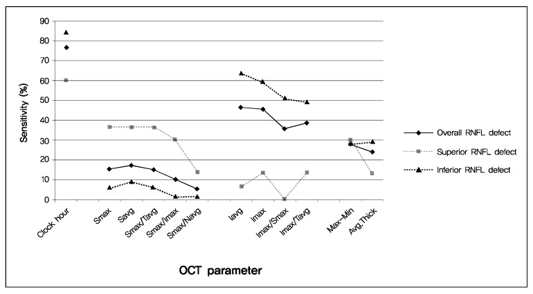
Figure 1 Graphical comparison of the specific table parameter according to the location of RNFL defect.
Reference
-
1. Sommer A, Miller NR, Pollack I, et al. The nerve fiber layer in the diagnosis of glaucoma. Arch ophthalmol. 1977. 95:2149–2156.2. Quigley HA, Addicks EM, Green WR. Optic nerve damage in human glaucoma. III. Quantitative correlation of nerve fiber loss and visual field defect in glaucoma, ischemic neuropathy, papilledema, and toxic neuropathy. Arch Ophthalmol. 1982. 100:135–146.3. Sommer A, Katz J, Quigley HA, et al. Clinically detectable nerve fiber atrophy precedes the onset of glaucomatous field loss. Arch Ophthalmol. 1991. 109:77–83.4. Quigley HA. Diagnosing Early glaucoma with Nerve Fiber layer Examination. 1996. 6th ed. New York: lgaku-Shoin;456–459.5. Lichter PR. Variability of expert observers in evaluating the optic disc. Trans Am Ophthalmol Soc. 1976. 74:532–572.6. Tielsch JM, Katz J, Quigley HA, et al. Intraobserver and interobserver agreement in measurement of optic disc characteristics. Ophthalmology. 1988. 95:350–356.7. Varma R, Steinmann WC, Scott IU. Expert agreement in evaluating the optic disc for glaucoma. Ophthalmology. 1992. 99:215–221.8. Jaffe GJ, Caprioli J. Optical coherence tomography to detect and manage retinal disease and glaucoma. Am J Ophthalmol. 2004. 137:156–169.9. Kim TW, Park UC, Park KH, Kim DM. Ability of stratus OCT to identify localized retinal nerve fiber layer defects in patients with normal standard automated perimetry results. Invest Ophthalmol Vis Sci. 2007. 48:1635–1641.10. Jeoung JW, Park KH, Kim TW, et al. Diagnostic ability of optical coherence tomography with a normative database to detect localized retinal nerve fiber layer defects. Ophthalmology. 2005. 112:2157–2163.11. Lu AT, Wang M, Varma R, et al. Combining nerve fiber layer parameters to optimize glaucoma diagnosis with optical coherence tomography. Ophthalmology. 2008. 115:1352–1357.12. Budenz DL, Michael A, Chang RT, et al. Sensitivity and specificity of the stratus OCT for perimetric glaucoma. Ophthalmology. 2005. 112:3–9.13. Hougaard JL, Heijl A, Bengtsson B. Glaucoma detection by stratus OCT. J Glaucoma. 2007. 16:302–306.14. Polo V, Larrosa JM, Ferreras A, et al. Retinal nerve fiber layer evaluation in open angle glaucoma. Ophthalmologica. 2009. 223:2–6.15. Paunescu LA, Schuman JS, Price LL, et al. Reproducibility of nerve fiber thickness, macular thickness, and optic nerve head measurements using Stratus OCT. Invest Ophthalmol Vis Sci. 2004. 45:1716–1724.16. Schuman JS, Pedut-Kloizman T, Hertzmark E, et al. Reproducibility of nerve fiber layer thickness measurements using optical coherence tomography. Ophthalmology. 1996. 103:1889–1898.17. Weinreb RN, Shakiba S, Zangwill L. Scanning laser polarimetry to measure the nerve fiber of normal and glaucomatous eyes. Am J Ophthalmol. 1995. 119:627–636.18. Guedes V, Schuman JS, Hertzmark E, et al. Optical coherence tomography measurement of macular and nerve fiber layer thickness in normal and glaucomatous human eyes. Ophthalmology. 2003. 110:177–189.19. Medeiros FA, Zangwill LM, Bowd C, et al. Fourier analysis of scanning laser polarimetry measurements with variable corneal compensation in glaucoma. Invest Ophthalmol Vis Sci. 2003. 44:2606–2612.20. Schuman JS, Hee MR, Puliafito CA, et al. Quantification of nerve fiber layer thickness in normal and glaucomatous eyes using optical coherence tomography. Arch Ophthalmol. 1995. 113:586–596.21. Hwang JM, Kim TW, Park KH, et al. Correlation between topographic profiles of localized retinal nerve fiber layer defects as determined by optical coherence tomography and red-free fundus photography. J Glaucoma. 2006. 15:223–228.22. Teesalu P, Tuulonen A, Airaksinen PJ. Optical coherence tomography and localized defects of the retinal nerve fiber layer. Acta Ophthalmol Scand. 2000. 78:49–52.23. Pieroth L, Schuman JS, Hertzmark E, et al. Evaluation of focal defects of the nerve fiber layer using optical coherence tomography. Ophthalmology. 1999. 106:570–579.24. Chen HY, Huang ML. Discrimination between normal and glaucomatous eyes using Stratus optical coherence tomography in Taiwan Chinese subjects. Graefes Arch Clin Exp Ophthalmol. 2005. 243:894–902.25. Huang ML, Chen HY. Development and comparison of automated classifiers for glaucoma diagnosis using stratus optical coherence tomography. Invest Ophthalmol Vis Sci. 2005. 46:4121–4129.26. Parikh RS, Parikh S, Sekhar GC, et al. Diagnostic capability of optical coherence tomography (stratus OCT3) in early glaucoma. Ophthalmology. 2007. 114:2238–2243.27. Bowd C, Zangwill LM, Berry CC, et al. Detecting early glaucoma by assessment of retinal nerve fiber layer thickness and visual function. Invest Ophthalmol Vis Sci. 2001. 42:1993–2003.28. Zangwill LM, Bowd LM, Berry CC, et al. Discriminating between normal and glaucomatous eyes using the Heidelberg retina tomograph, GDx nerve fiber analyzer, and optical coherence tomograph. Arch Ophthalmol. 2001. 119:985–993.29. Kamal DS, Garway-Heath DF, Hitchings RA, Fitzke FW. Use of sequential Heidelberg retina tomograph images to identify changes at the optic disc in ocular hypertensive patients at risk of developing glaucoma. Br J Ophthalmol. 2000. 84:993–998.30. Parisi V, Manni G, Centofanti M, et al. Correlation between optical coherence tomography, pattern electroretinogram, and visual evoked potentials in open-angle glaucoma patients. Ophthalmology. 2001. 108:905–912.31. Medeiros FA, Zangwill LM, Bowd C, et al. Comparison of scanning laser polarimetry using variable corneal compensation and retinal nerve fiber layer photography for detection of glaucoma. Arch Ophthalmol. 2004. 122:698–704.32. Vernon SA, Rotchford AP, Negi A, et al. Peripapillary retinal nerve fibre layer thickness in highly myopic Caucasians as measured by Stratus optical coherence tomography. Br J Ophthalmol. 2008. 92:1076–1080.33. Leung CK, Mohamed S, Leung KS, et al. Retinal nerve fiber layer measurements in myopia: An optical coherence tomography study. Invest Ophthalmol Vis Sci. 2006. 47:5171–5176.34. Kanamori A, Nakamura M, Escano MF, et al. Evaluation of the glaucomatous damage on retinal nerve fiber layer thickness measured by optical coherence tomography. Am J Ophthalmol. 2003. 135:513–520.35. Caprioli J. The contour of the juxtapapillary nerve fiber layer in glaucoma. Ophthalmology. 1990. 97:358–365.36. Leung CK, Chan WM, Yung WH, et al. Comparison of macular and peripapillary measurements for the detection of glaucoma: an optical coherence tomography study. Ophthalmology. 2005. 112:391–400.37. Budenz DL, Anderson DR, Varma R, et al. Deternimants of normal retinal nerve fiber layer thickness measured by stratus OCT. Ophthalmology. 2007. 114:1046–1052.38. Hougaard JL, Heijl A, Bengtsson B. Glaucoma detection using different stratus optical coherence tomography protocols. Acta Ophthalmol Scand. 2007. 85:251–256.39. Da pozzo S, Iacono P, Marchesan R, et al. The effect of aging on retinal nerve fibre layer thickness: an evaluation by scanning laser polarimetry with variable corneal compensation. Acta Ophthalmol Scand. 2006. 84:375–379.40. Song TG, Yoo YC, Lee HB. Quantitative analysis of retinal nerve fiber layer thickness profile in myopic eyes. J Korean Ophthalmol Soc. 2009. 50:1840–1846.
- Full Text Links
-
- Actions
-
Cited
- CITED
-
- Close
- Share
-
- Similar articles
-
- Comparison of Time Domain OCT and Spectrum Domain OCT for Retinal Nerve Fiber Layer Assessment
- Comparison of Diagnostic Ability of 3D and Stratus Optical Coherence Tomography in Early Glaucoma
- Diagnostic Ability of Stratus OCT Using Korean Normative Database for Early Detection of Normal-Tension Glaucoma
- Comparison of the Results between Heidelberg Retina Tomography II and Stratus Optical Coherence Tomography in Glaucoma
- Comparison of Stratus OCT and GDx VCC in Detecting Localized Retinal Nerve Fiber Layer Defects
