J Adv Prosthodont.
2014 Jun;6(3):200-206. 10.4047/jap.2014.6.3.200.
Fracture resistance of endodontically treated maxillary premolars restored by silorane-based composite with or without fiber or nano-ionomer
- Affiliations
-
- 1Department of Operative Dentistry, School of Dentistry, Shiraz University of Medical Sciences, Shiraz, Iran. tavangarm@yahoo.com
- 2Biomaterial Research Center, School of Dentistry, Shiraz University of Medical Sciences, Shiraz, Iran.
- 3Department of Endodontics, School of Dentistry, Shiraz University of Medical Sciences, Shiraz, Iran.
- KMID: 2118231
- DOI: http://doi.org/10.4047/jap.2014.6.3.200
Abstract
- PURPOSE
This in vitro study investigated the fracture resistance of endodontically treated premolars restored using silorane- or methacrylate-based composite along with or without fiber or nano-ionomer base.
MATERIALS AND METHODS
Ninety-six intact maxillary premolars were randomly divided into eight groups (n = 12). G1 (negative control) was the intact teeth. In Groups 2-8, root canal treatment with mesio-occlusodistal preparation was performed. G2 (positive control) was kept unrestored. The other groups were restored using composite resin as follows: G3, methacrylate-based composite (Z250); G4, methacrylate composite (Z250) with polyethylene fiber; G5 and G6, silorane-based composite (Filtek P90) without and with the fiber, respectively; G7 and G8, methacrylate- and silorane-based composite with nano-ionomer base, respectively. After aging period and thermocycling for 1000 cycles, fracture strength was tested and fracture patterns were inspected. The results were analyzed using ANOVA and Tukey HSD tests (alpha=0.05).
RESULTS
Mean fracture resistance for the eight groups (in Newton) were G1: 1200 +/- 169a, G2: 360 +/- 93b, G3: 632 +/- 196c, G4: 692 +/- 195c, G5: 917 +/- 159d, G6: 1013 +/- 125ad, G7: 959 +/- 148d, G8: 947 +/- 105d (different superscript letters revealed significant difference among groups). Most of the fractures in all the groups were restorable, except Group 3.
CONCLUSION
Silorane-based composite revealed significantly higher strength of the restored premolars compared to that of methacrylate one. Fiber insertion demonstrated no additional effect on the strength of both composite restorations; however, it increased the prevalence of restorable fracture of methacrylate-based composite restored teeth. Using nano-ionomer base under methacrylate-based composite had a positive effect on fracture resistance and pattern. Only fiber-reinforced silorane composite restoration resulted in a strength similar to that of the intact teeth.
Keyword
MeSH Terms
Figure
-
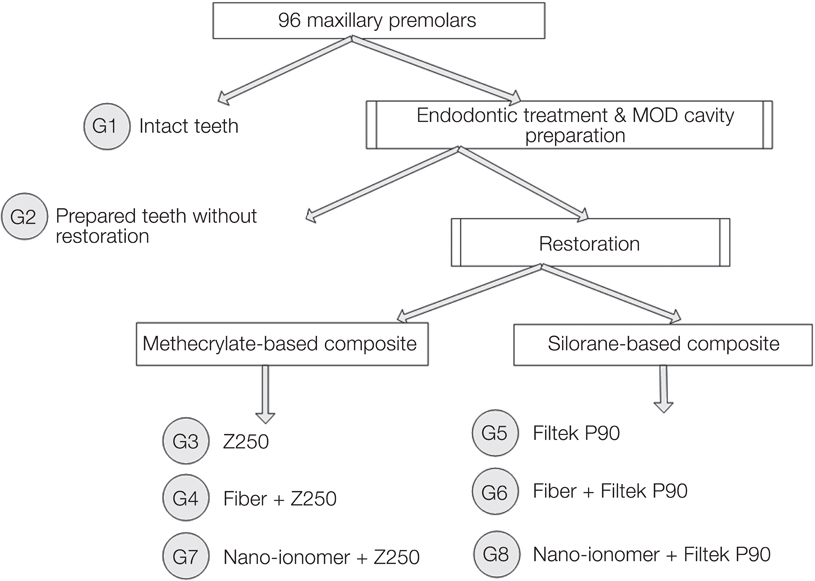
Fig. 1 Descriptive diagram of the eight groups.

Fig. 2 Mode of restorable fracture.
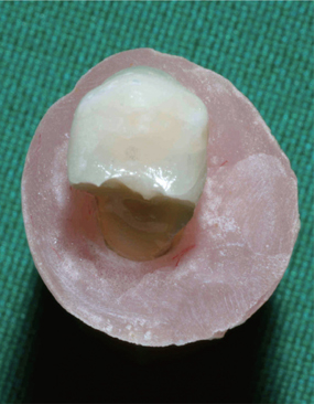
Fig. 3 Mode of unrestorable fracture.
Reference
-
1. Steele A, Johnson BR. In vitro fracture strength of endodontically treated premolars. J Endod. 1999; 25:6–8.2. Krejci I, Duc O, Dietschi D, de Campos E. Marginal adaptation, retention and fracture resistance of adhesive composite restorations on devital teeth with and without posts. Oper Dent. 2003; 28:127–135.3. Trope M, Langer I, Maltz D, Tronstad L. Resistance to fracture of restored endodontically treated premolars. Endod Dent Traumatol. 1986; 2:35–38.4. Soares PV, Santos-Filho PC, Queiroz EC, Araújo TC, Campos RE, Araújo CA, Soares CJ. Fracture resistance and stress distribution in endodontically treated maxillary premolars restored with composite resin. J Prosthodont. 2008; 17:114–119.5. Taha NA, Palamara JE, Messer HH. Cuspal deflection, strain and microleakage of endodontically treated premolar teeth restored with direct resin composites. J Dent. 2009; 37:724–730.6. Rodrigues FB, Paranhos MP, Spohr AM, Oshima HM, Carlini B, Burnett LH Jr. Fracture resistance of root filled molar teeth restored with glass fibre bundles. Int Endod J. 2010; 43:356–362.7. Randow K, Glantz PO. On cantilever loading of vital and non-vital teeth. An experimental clinical study. Acta Odontol Scand. 1986; 44:271–277.8. de Freitas CR, Miranda MI, de Andrade MF, Flores VH, Vaz LG, Guimarães C. Resistance to maxillary premolar fractures after restoration of class II preparations with resin composite or ceromer. Quintessence Int. 2002; 33:589–594.9. Schwartz RS, Robbins JW. Post placement and restoration of endodontically treated teeth: a literature review. J Endod. 2004; 30:289–301.10. Soares PV, Santos-Filho PC, Martins LR, Soares CJ. Influence of restorative technique on the biomechanical behavior of endodontically treated maxillary premolars. Part I: fracture resistance and fracture mode. J Prosthet Dent. 2008; 99:30–37.11. Hürmüzlü F, Kiremitçi A, Serper A, Altundaşar E, Siso SH. Fracture resistance of endodontically treated premolars restored with ormocer and packable composite. J Endod. 2003; 29:838–840.12. Siso SH, Hürmüzlü F, Turgut M, Altundaşar E, Serper A, Er K. Fracture resistance of the buccal cusps of root filled maxillary premolar teeth restored with various techniques. Int Endod J. 2007; 40:161–168.13. Yamada Y, Tsubota Y, Fukushima S. Effect of restoration method on fracture resistance of endodontically treated maxillary premolars. Int J Prosthodont. 2004; 17:94–98.14. Mohammadi N, Kahnamoii MA, Yeganeh PK, Navimipour EJ. Effect of fiber post and cusp coverage on fracture resistance of endodontically treated maxillary premolars directly restored with composite resin. J Endod. 2009; 35:1428–1432.15. Scotti N, Scansetti M, Rota R, Pera F, Pasqualini D, Berutti E. The effect of the post length and cusp coverage on the cycling and static load of endodontically treated maxillary premolars. Clin Oral Investig. 2011; 15:923–929.16. Freedman GA. Esthetic post-and-core treatment. Dent Clin North Am. 2001; 45:103–116.17. Salameh Z, Sorrentino R, Papacchini F, Ounsi HF, Tashkandi E, Goracci C, Ferrari M. Fracture resistance and failure patterns of endodontically treated mandibular molars restored using resin composite with or without translucent glass fiber posts. J Endod. 2006; 32:752–755.18. Soares CJ, Soares PV, de Freitas Santos-Filho PC, Castro CG, Magalhaes D, Versluis A. The influence of cavity design and glass fiber posts on biomechanical behavior of endodontically treated premolars. J Endod. 2008; 34:1015–1019.19. Fokkinga WA, Le Bell AM, Kreulen CM, Lassila LV, Vallittu PK, Creugers NH. Ex vivo fracture resistance of direct resin composite complete crowns with and without posts on maxillary premolars. Int Endod J. 2005; 38:230–237.20. Bitter K, Meyer-Lueckel H, Fotiadis N, Blunck U, Neumann K, Kielbassa AM, Paris S. Influence of endodontic treatment, post insertion, and ceramic restoration on the fracture resistance of maxillary premolars. Int Endod J. 2010; 43:469–477.21. Eskitaşcioğlu G, Belli S, Kalkan M. Evaluation of two post core systems using two different methods (fracture strength test and a finite elemental stress analysis). J Endod. 2002; 28:629–633.22. Hitz T, Ozcan M, Göhring TN. Marginal adaptation and fracture resistance of root-canal treated mandibular molars with intracoronal restorations: effect of thermocycling and mechanical loading. J Adhes Dent. 2010; 12:279–286.23. Versluis A, Douglas WH, Cross M, Sakaguchi RL. Does an incremental filling technique reduce polymerization shrinkage stresses? J Dent Res. 1996; 75:871–878.24. Karbhari VM, Wang Q. Influence of triaxial braid denier on ribbon-based fiber reinforced dental composites. Dent Mater. 2007; 23:969–976.25. Belli S, Dönmez N, Eskitaşcioğlu G. The effect of c-factor and flowable resin or fiber use at the interface on microtensile bond strength to dentin. J Adhes Dent. 2006; 8:247–253.26. Belli S, Erdemir A, Yildirim C. Reinforcement effect of polyethylene fibre in root-filled teeth: comparison of two restoration techniques. Int Endod J. 2006; 39:136–142.27. Palin WM, Fleming GJ, Nathwani H, Burke FJ, Randall RC. In vitro cuspal deflection and microleakage of maxillary premolars restored with novel low-shrink dental composites. Dent Mater. 2005; 21:324–335.28. Taha NA, Palamara JE, Messer HH. Fracture strength and fracture patterns of root filled teeth restored with direct resin restorations. J Dent. 2011; 39:527–535.29. Abd El Halim S, Zaki D. Comparative evaluation of microleakage among three different glass ionomer types. Oper Dent. 2011; 36:36–42.30. Shafiei F, Akbarian S. Microleakage of nanofilled resin-modified glass-ionomer/silorane- or methacrylate-based composite sandwich Class II restoration: effect of simultaneous bonding. Oper Dent. 2014; 39:E22–E30.31. Uyehara MY, Davis RD, Overton JD. Cuspal reinforcement in endodontically treated molars. Oper Dent. 1999; 24:364–370.32. Sengun A, Cobankara FK, Orucoglu H. Effect of a new restoration technique on fracture resistance of endodontically treated teeth. Dent Traumatol. 2008; 24:214–219.33. Cobankara FK, Unlu N, Cetin AR, Ozkan HB. The effect of different restoration techniques on the fracture resistance of endodontically-treated molars. Oper Dent. 2008; 33:526–533.34. Belli S, Erdemir A, Ozcopur M, Eskitascioglu G. The effect of fibre insertion on fracture resistance of root filled molar teeth with MOD preparations restored with composite. Int Endod J. 2005; 38:73–80.35. Oskoee PA, Ajami AA, Navimipour EJ, Oskoee SS, Sadjadi J. The effect of three composite fiber insertion techniques on fracture resistance of root-filled teeth. J Endod. 2009; 35:413–416.36. González-López S, Lucena-Martín C, de Haro-Gasquet F, Vilchez-Díaz MA, de Haro-Muñoz C. Influence of different composite restoration techniques on cuspal deflection: an in vitro study. Oper Dent. 2004; 29:656–660.37. Jafarpour S, El-Badrawy W, Jazi HS, McComb D. Effect of composite insertion technique on cuspal deflection using an in vitro simulation model. Oper Dent. 2012; 37:299–305.38. Karaman E, Ozgunaltay G. Cuspal deflection in premolar teeth restored using current composite resins with and without resin-modified glass ionomer liner. Oper Dent. 2013; 38:282–289.39. Agrawal VS, Parekh VV, Shah NC. Comparative evaluation of microleakage of silorane-based composite and nanohybrid composite with or without polyethylene fiber inserts in class II restorations: an in vitro study. Oper Dent. 2012; 37:e1–e7.40. Buergers R, Schneider-Brachert W, Hahnel S, Rosentritt M, Handel G. Streptococcal adhesion to novel low-shrink silorane-based restorative. Dent Mater. 2009; 25:269–275.41. Xie KX, Wang XY, Gao XJ, Yuan CY, Li JX, Chu CH. Fracture resistance of root filled premolar teeth restored with direct composite resin with or without cusp coverage. Int Endod J. 2012; 45:524–529.42. Shafiei F, Memarpour M, Karimi F. Fracture resistance of cuspal coverage of endodontically treated maxillary premolars with combined composite-amalgam compared to other techniques. Oper Dent. 2011; 36:439–447.
- Full Text Links
-
- Actions
-
Cited
- CITED
-
- Close
- Share
-
- Similar articles
-
- Fracture resistance of upper central incisors restored with different posts and cores
- A comparative evaluation of fracture resistance of endodontically treated teeth restored with different post core systems: an in-vitro study
- Stress distribution of endodontically treated maxillary second premolars restored with different methods: Three-dimensional finite element analysis
- Fracture resistance of endodontically treated canines restored with different sizes of fiber post and all-ceramic crowns
- Fracture resistances of zirconia, cast Ni-Cr, and fiber-glass composite posts under all-ceramic crowns in endodontically treated premolars
