Clinical gap changes after porcelain firing cycles of zirconia fixed dentures
- Affiliations
-
- 1Private Dentist, Istanbul, Turkey.
- 2Department of Prosthodontics, Faculty of Dentistry, Marmara University, Istanbul, Turkey. begumturker@hotmail.com
- KMID: 2118228
- DOI: http://doi.org/10.4047/jap.2014.6.3.177
Abstract
- PURPOSE
The aim of this study was to measure the changes on the marginal and internal adaptation of zirconia based anterior fixed partial dentures after the porcelain firing process.
MATERIALS AND METHODS
A total of 34 anterior fixed partial dentures using LAVA CAD/CAM system (3M ESPE, Germany) were applied. Two silicone replicas were obtained: one is obtained before porcelain firing process (initial) and the other is obtained after porcelain firing process (final), followed by the examination under a binocular stereomicroscope. Kruskal Wallis and Wilcoxon Signed Ranks tests were used for the statistical analysis (P<.05).
RESULTS
No statistically significant difference was found between initial and final marginal gap values (P>.05). At the internal gap measurements, final marginal area values (59.54 microm) were significantly lower than the initial marginal area values (68.68 microm)(P<.05). The highest and the lowest internal gap values were observed at the incisal/occlusal area and at the marginal area, respectively. In addition, lower internal gap values were obtained for canines than for central incisors, lateral incisors and premolars at the incisal area (P<.05).
CONCLUSION
The firing cycles did not affect the marginal gap of Lava CAD/CAM system, but it is controversial for the internal gap.
Keyword
MeSH Terms
Figure
-
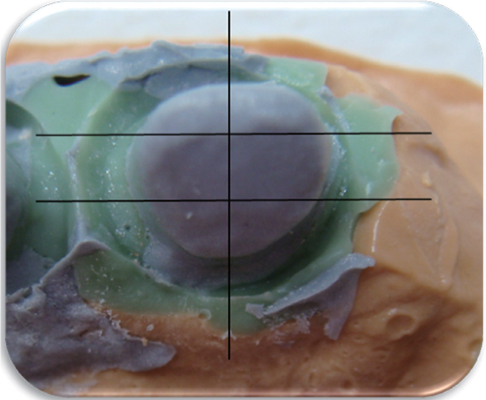
Fig. 1 Silicone replica. Black lines indicate the intersections for specimens preparation.
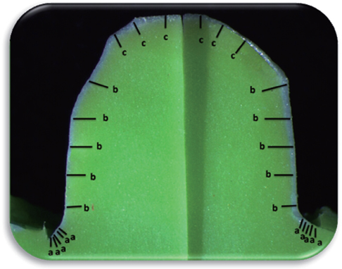
Fig. 2 Stereomicroscope image of a transversal slice. a: Marginal gap measurement points, b: Axial gap measurement points, c: Icisal gap measurement points.
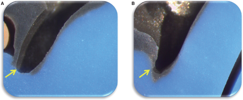
Fig. 3 (A) Marginal adaptation Type: 1 (No marginal gap), (B) Marginal adaptation Type: 2 (Marginal gap: 54.64 µm, no overcontour or undercontour). Yellow arrow indicates marginal adaptation.

Fig. 4 (A) Initial stereomicroscope image of marginal area (mean gap value: 85.16 µm), (B) Final stereomicroscope image of marginal area (mean gap value: 64.76 µm). Yellow arrow indicates the marginal gap area.
Cited by 1 articles
-
Verification of a computer-aided replica technique for evaluating prosthesis adaptation using statistical agreement analysis
Hang-Nga Mai, Kyeong Eun Lee, Kyu-Bok Lee, Seung-Mi Jeong, Seok-Jae Lee, Cheong-Hee Lee, Seo-Young An, Du-Hyeong Lee
J Adv Prosthodont. 2017;9(5):358-363. doi: 10.4047/jap.2017.9.5.358.
Reference
-
1. Raigrodski AJ. Contemporary materials and technologies for all-ceramic fixed partial dentures: a review of the literature. J Prosthet Dent. 2004; 92:557–562.2. Aboushelib MN, de Jager N, Kleverlaan CJ, Feilzer AJ. Microtensile bond strength of different components of core veneered all-ceramic restorations. Dent Mater. 2005; 21:984–991.3. Yanagida H, Kōmoto K, Miyayama M. The chemistry of ceramics. Chichester; New York: Wiley;1996.4. Piwowarczyk A, Ottl P, Lauer HC, Kuretzky T. A clinical report and overview of scientific studies and clinical procedures conducted on the 3M ESPE Lava All-Ceramic System. J Prosthodont. 2005; 14:39–45.5. Holmes JR, Bayne SC, Holland GA, Sulik WD. Considerations in measurement of marginal fit. J Prosthet Dent. 1989; 62:405–408.6. Sulaiman F, Chai J, Jameson LM, Wozniak WT. A comparison of the marginal fit of In-Ceram, IPS Empress, and Procera crowns. Int J Prosthodont. 1997; 10:478–484.7. Jacobs MS, Windeler AS. An investigation of dental luting cement solubility as a function of the marginal gap. J Prosthet Dent. 1991; 65:436–442.8. Goldman M, Laosonthorn P, White RR. Microleakage-full crowns and the dental pulp. J Endod. 1992; 18:473–475.9. Jørgensen KD. Factors affecting the film thickness of zinc phosphate cements. Acta Odontol Scand. 1960; 18:479–490.10. Tinschert J, Natt G, Mautsch W, Spiekermann H, Anusavice KJ. Marginal fit of alumina-and zirconia-based fixed partial dentures produced by a CAD/CAM system. Oper Dent. 2001; 26:367–374.11. Boening KW, Wolf BH, Schmidt AE, Kästner K, Walter MH. Clinical fit of Procera AllCeram crowns. J Prosthet Dent. 2000; 84:419–424.12. Reich S, Wichmann M, Nkenke E, Proeschel P. Clinical fit of all-ceramic three-unit fixed partial dentures, generated with three different CAD/CAM systems. Eur J Oral Sci. 2005; 113:174–179.13. Reich S, Kappe K, Teschner H, Schmitt J. Clinical fit of four-unit zirconia posterior fixed dental prostheses. Eur J Oral Sci. 2008; 116:579–584.14. Gonzalo E, Suárez MJ, Serrano B, Lozano JF. Marginal fit of Zirconia posterior fixed partial dentures. Int J Prosthodont. 2008; 21:398–399.15. Vigolo P, Fonzi F. An in vitro evaluation of fit of zirconium-oxide-based ceramic four-unit fixed partial dentures, generated with three different CAD/CAM systems, before and after porcelain firing cycles and after glaze cycles. J Prosthodont. 2008; 17:621–626.16. Gonzalo E, Suárez MJ, Serrano B, Lozano JF. A comparison of the marginal vertical discrepancies of zirconium and metal ceramic posterior fixed dental prostheses before and after cementation. J Prosthet Dent. 2009; 102:378–384.17. Beuer F, Aggstaller H, Edelhoff D, Gernet W, Sorensen J. Marginal and internal fits of fixed dental prostheses zirconia retainers. Dent Mater. 2009; 25:94–102.18. Wettstein F, Sailer I, Roos M, Hämmerle CH. Clinical study of the internal gaps of zirconia and metal frameworks for fixed partial dentures. Eur J Oral Sci. 2008; 116:272–279.19. Denissen H, Dozić A, van der Zel J, van Waas M. Marginal fit and short-term clinical performance of porcelain-veneered CICERO, CEREC, and Procera onlays. J Prosthet Dent. 2000; 84:506–513.20. Bindl A, Mörmann WH. Marginal and internal fit of all-ceramic CAD/CAM crown-copings on chamfer preparations. J Oral Rehabil. 2005; 32:441–447.21. Reich S, Uhlen S, Gozdowski S, Lohbauer U. Measurement of cement thickness under lithium disilicate crowns using an impression material technique. Clin Oral Investig. 2011; 15:521–526.22. Kokubo Y, Nagayama Y, Tsumita M, Ohkubo C, Fukushima S, Vult von Steyern P. Clinical marginal and internal gaps of In-Ceram crowns fabricated using the GN-I system. J Oral Rehabil. 2005; 32:753–758.23. Castellani D, Baccetti T, Clauser C, Bernardini UD. Thermal distortion of different materials in crown construction. J Prosthet Dent. 1994; 72:360–366.24. Balkaya MC, Cinar A, Pamuk S. Influence of firing cycles on the margin distortion of 3 all-ceramic crown systems. J Prosthet Dent. 2005; 93:346–355.25. Att W, Komine F, Gerds T, Strub JR. Marginal adaptation of three different zirconium dioxide three-unit fixed dental prostheses. J Prosthet Dent. 2009; 101:239–247.26. Dittmer MP, Borchers L, Stiesch M, Kohorst P. Stresses and distortions within zirconia-fixed dental prostheses due to the veneering process. Acta Biomater. 2009; 5:3231–3239.27. Kohorst P, Brinkmann H, Dittmer MP, Borchers L, Stiesch M. Influence of the veneering process on the marginal fit of zirconia fixed dental prostheses. J Oral Rehabil. 2010; 37:283–291.28. Aboushelib MN, Feilzer AJ, de Jager N, Kleverlaan CJ. Prestresses in bilayered all-ceramic restorations. J Biomed Mater Res B Appl Biomater. 2008; 87:139–145.29. Pospiech PR, Rountree PR, Nothdurft FP. Clinical evaluation of zirconia-based all ceramic posterior bridges: two-year results. J Dent Res. 2003; 82:114.30. Raigrodski AJ, Chiche GJ, Potiket N, Hochstedler JL, Mohamed SE, Billiot S, Mercante DE. The efficacy of posterior three-unit zirconium-oxide-based ceramic fixed partial dental prostheses: a prospective clinical pilot study. J Prosthet Dent. 2006; 96:237–244.31. Crisp RJ, Cowan AJ, Lamb J, Thompson O, Tulloch N, Burke FJ. A clinical evaluation of all-ceramic bridges placed in UK general dental practices: first-year results. Br Dent J. 2008; 205:477–482.
- Full Text Links
-
- Actions
-
Cited
- CITED
-
- Close
- Share
-
- Similar articles
-
- The effects of repetitive firing processes on the optical, thermal, and phase formation changes of zirconia
- Study of the fracture resistance of zirconia on posterior fixed partial dentures based on inter-abutment distance
- A comparison of marginal fit of glass infiltrated alumina copings fabricated using two different techniques and the effect of firing cycles over them
- Impact of multiple firings and resin cement type on shear bond strength between zirconia and resin cements
- Implant supported fixed prosthesis for complete edentulous maxilla with severe alveolar ridge resorption: A case report
