Effect of polishing and glazing on the color and spectral distribution of monolithic zirconia
- Affiliations
-
- 1Department of Prosthodontics and Dental Research Institute, School of Dentistry, Seoul National University, Seoul, Republic of Korea. swallow@snu.ac.kr
- KMID: 2118169
- DOI: http://doi.org/10.4047/jap.2013.5.3.296
Abstract
- PURPOSE
The aim of this study was to evaluate the effect of polishing and glazing on the color and spectral distribution of monolithic zirconia.
MATERIALS AND METHODS
Forty-five monolithic zirconia specimens (16.3 mm x 16.4 mm x 2.0 mm) were fabricated and divided into 5 groups according to the number of A2-coloring liquid applications (Group I to V). Each group was divided into 3 subgroups according to the method of surface treatments (n=3): N: no treatment; P: polishing; G: glazing. Color and spectral distribution of five different areas of each specimen were measured according to CIELAB color space in the reflectance mode relative to the standard illuminant D65 on a reflection spectrophotometer. Data were analyzed using one-way ANOVA followed by Tukey's HSD test, Pearson correlation and regression analysis (alpha=.05).
RESULTS
There was a significant difference in CIE L* between Subgroup N and P, and in CIE b* between Subgroup P and G in each group. Spectral reflectance generally decreased in Subgroup P and G in comparison with Subgroup N. Color differences between Subgroup P and G were within the perceptibility threshold (DeltaE*ab< 3.7) in most groups. Highly significant correlation was found between CIE b*and each subgroups as the number of coloring liquid applications increased (R2>0.88, P<.001).
CONCLUSION
A perceptible color difference can be detected after polishing of monolithic zirconia. Polishing decreases the lightness, and glazing also decreases the lightness, but increases the yellowness of monolithic zirconia.
Figure
-
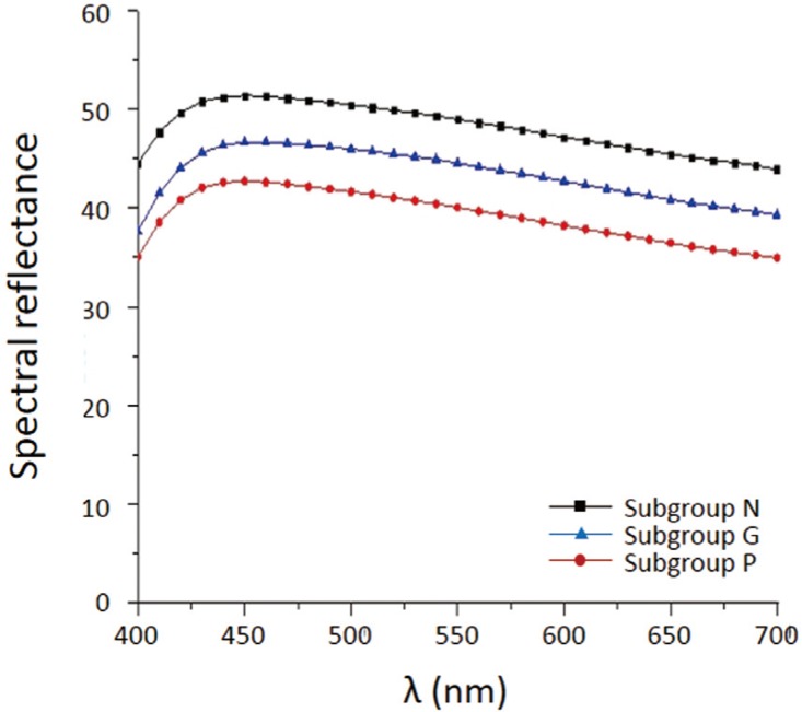
Fig. 1 Spectral reflectance of each subgroup in Group I against white background.
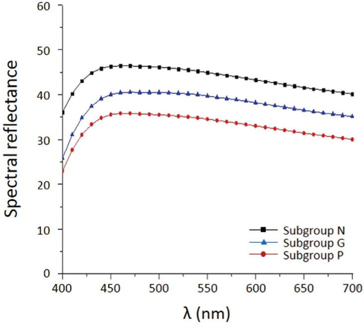
Fig. 2 Spectral reflectance of each subgroup in Group II against white background.
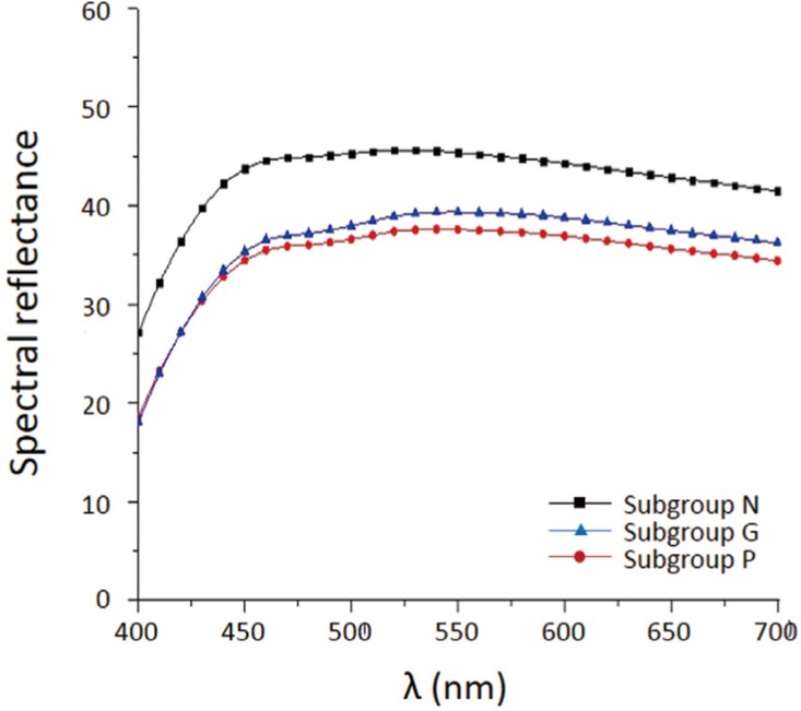
Fig. 3 Spectral reflectance of each subgroup in Group III against white background.
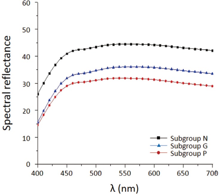
Fig. 4 Spectral reflectance of each subgroup in Group IV against white background.
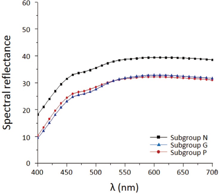
Fig. 5 Spectral reflectance of each subgroup in Group V against white background.
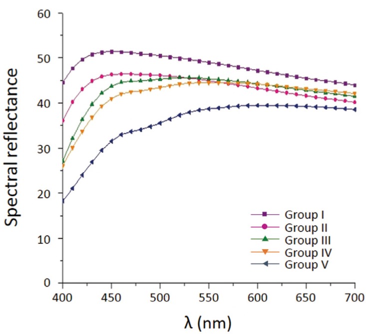
Fig. 6 Spectral reflectance of each group within Subgroup N.
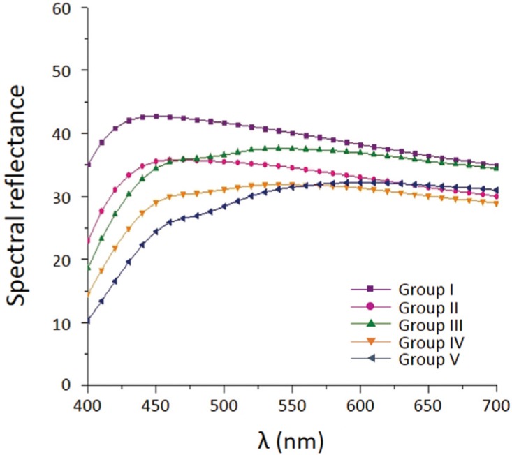
Fig. 7 Spectral reflectance of each group within Subgroup P.
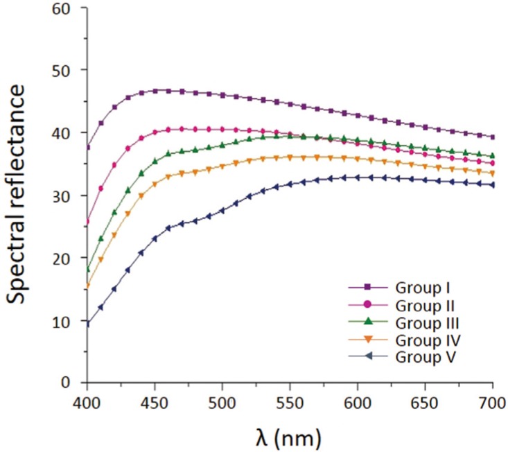
Fig. 8 Spectral reflectance of each group within Subgroup G.
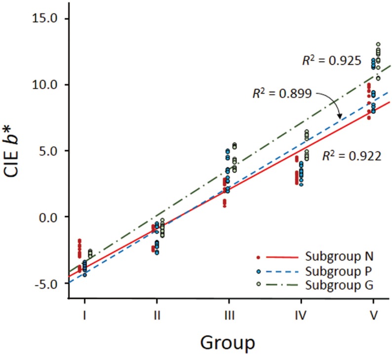
Fig. 9 Linear regression of CIE b* values of each subgroup over a zero calibration box in the reflectance mode as a function of the number of coloring liquid applications.
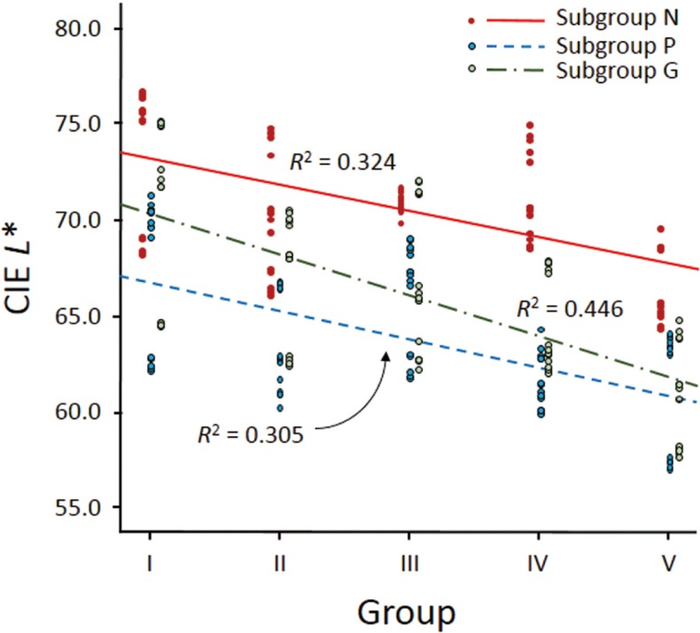
Fig. 10 Linear regression of CIE L* values of each subgroup over a zero calibration box in the reflectance mode as a function of the number of coloring liquid applications.
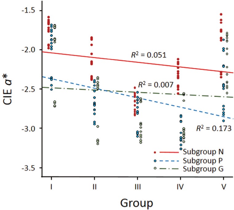
Fig. 11 Linear regression of CIE a* values of each subgroup over a zero calibration box in the reflectance mode as a function of the number of coloring liquid applications.
Reference
-
1. Weinstein M, Katz S, Weinstein AB. Fused porcelain-to-metal teeth. US patent No 3052, 982. 1962.2. Pröbster L, Diehl J. Slip-casting alumina ceramics for crown and bridge restorations. Quintessence Int. 1992; 23:25–31. PMID: 1631267.3. Vult von Steyern P, Carlson P, Nilner K. All-ceramic fixed partial dentures designed according to the DC-Zirkon technique. A 2-year clinical study. J Oral Rehabil. 2005; 32:180–187. PMID: 15707428.4. Tinschert J, Natt G, Mautsch W, Augthun M, Spiekermann H. Fracture resistance of lithium disilicate-, alumina-, and zirconia-based three-unit fixed partial dentures: a laboratory study. Int J Prosthodont. 2001; 14:231–238. PMID: 11484570.5. Al-Amleh B, Lyons K, Swain M. Clinical trials in zirconia: a systematic review. J Oral Rehabil. 2010; 37:641–652. PMID: 20406352.
Article6. Ha SR, Kim SH, Han JS, Yoo SH, Jeong SC, Lee JB, Yeo IS. The influence of various core designs on stress distribution in the veneered zirconia crown: a finite element analysis study. J Adv Prosthodont. 2013; 5:187–197. PMID: 23755346.
Article7. Beuer F, Schweiger J, Eichberger M, Kappert HF, Gernet W, Edelhoff D. High-strength CAD/CAM-fabricated veneering material sintered to zirconia copings-a new fabrication mode for all-ceramic restorations. Dent Mater. 2009; 25:121–128. PMID: 18620748.8. Aboushelib MN, Kleverlaan CJ, Feilzer AJ. Microtensile bond strength of different components of core veneered all-ceramic restorations. Part II: Zirconia veneering ceramics. Dent Mater. 2006; 22:857–863. PMID: 16376981.9. Aboushelib MN, Kleverlaan CJ, Feilzer AJ. Microtensile bond strength of different components of core veneered all-ceramic restorations. Part 3: double veneer technique. J Prosthodont. 2008; 17:9–13. PMID: 17931369.
Article10. Wiskott HWA. Fixed prosthodontics: principles and clinics. London: Quintessence publishing Co. Ltd;2011. p. 670–671.11. Klausner LH, Cartwright CB, Charbeneau GT. Polished versus autoglazed porcelain surfaces. J Prosthet Dent. 1982; 47:157–162. PMID: 7035652.
Article12. Brewer JD, Garlapo DA, Chipps EA, Tedesco LA. Clinical discrimination between autoglazed and polished porcelain surfaces. J Prosthet Dent. 1990; 64:631–634. PMID: 2079666.
Article13. Scurria MS, Powers JM. Surface roughness of two polished ceramic materials. J Prosthet Dent. 1994; 71:174–177. PMID: 8126673.
Article14. Kim IJ, Lee YK, Lim BS, Kim CW. Effect of surface topography on the color of dental porcelain. J Mater Sci Mater Med. 2003; 14:405–409. PMID: 15348443.15. Sarac D, Sarac YS, Yuzbasioglu E, Bal S. The effects of porcelain polishing systems on the color and surface texture of feldspathic porcelain. J Prosthet Dent. 2006; 96:122–128. PMID: 16911889.
Article16. Yilmaz C, Korkmaz T, Demirköprülü H, Ergün G, Ozkan Y. Color stability of glazed and polished dental porcelains. J Prosthodont. 2008; 17:20–24. PMID: 17971115.17. Commission Internationale de l'Eclairage (CIE). Colorimetry, CIE 015. 3rd ed. Vienna: CIE Central Bureau;2004.18. Obregon A, Goodkind RJ, Schwabacher WB. Effects of opaque and porcelain surface texture on the color of ceramometal restorations. J Prosthet Dent. 1981; 46:330–340. PMID: 7021809.
Article19. Lee YK, Lim BS, Kim CW. Effect of surface conditions on the color of dental resin composites. J Biomed Mater Res. 2002; 63:657–663. PMID: 12209913.
Article20. Chung KH. Effects of finishing and polishing procedures on the surface texture of resin composites. Dent Mater. 1994; 10:325–330. PMID: 7498594.
Article21. Knispel G. Factors affecting the process of color matching restorative materials to natural teeth. Quintessence Int. 1991; 22:525–531. PMID: 1882045.22. Heffernan MJ, Aquilino SA, Diaz-Arnold AM, Haselton DR, Stanford CM, Vargas MA. Relative translucency of six all-ceramic systems. Part I: core materials. J Prosthet Dent. 2002; 88:4–9. PMID: 12239472.
Article23. Baldissara P, Llukacej A, Ciocca L, Valandro FL, Scotti R. Translucency of zirconia copings made with different CAD/CAM systems. J Prosthet Dent. 2010; 104:6–12. PMID: 20620365.
Article24. Rosenstiel SF, Baiker MA, Johnston WM. Comparison of glazed and polished dental porcelain. Int J Prosthodont. 1989; 2:524–529. PMID: 2640126.
- Full Text Links
-
- Actions
-
Cited
- CITED
-
- Close
- Share
-
- Similar articles
-
- The effect of various polishing systems on surface roughness and phase transformation of monolithic zirconia
- Review on factors affecting the optical properties of dental zirconia
- The metameric effect of monolithic zirconias with varying yttrium ratios
- Effect of surface finishing treatments on the color stability of CAD/CAM materials
- Esthetic anterior restoration using 3M Lavaâ„¢ Esthetic monolithic zirconia
