Endoscopic Endonasal Treatment of a Pott's Puffy Tumor
- Affiliations
-
- 1Department of Otorhinolaryngology-Head and Neck Surgery, Korea University College of Medicine, Seoul, Korea. lhman@korea.ac.kr
- KMID: 2117506
- DOI: http://doi.org/10.3342/ceo.2012.5.2.112
Abstract
- Pott's puffy tumor is an infrequent entity characterized by a subperiosteal abscess associated with frontal bone osteomyelitis. It has become rare due to the development of antibiotics and is usually seen as a complication of frontal sinusitis. Although Pott's puffy tumor is more commonly described in children, it should also be included in the differential diagnosis of swelling on the forehead in adults. Once the diagnosis is suspected, appropriate imaging should be performed to evaluate the possible complications. The treatment of Pott's puffy tumor combines medical and surgical approaches in order to prevent further complications. The goal of surgery is to drain the sinus and to excise the infected bone if necessary. The endoscopic endonasal approach is a safe and effective alternative to the external approach. This report describes the case of a 25-year-old man with Pott's puffy tumor resulting from frontal sinusitis.
Keyword
MeSH Terms
Figure
-

Fig. 1 Patient profile showing the soft tissue swelling of the left forehead.
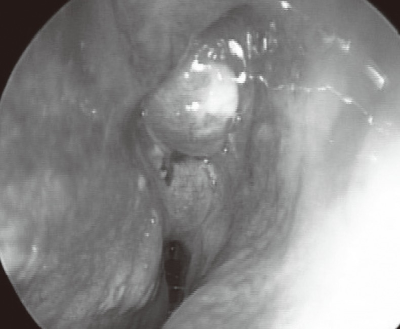
Fig. 2 Nasal endoscopic examination showing nasal polyp and purulent discharge in the right nasal cavity.

Fig. 3 (A) Axial CT showing left and right frontal sinusitis with right anterior bone defect (arrow). (B) Coronal CT showing both ethmoid and left maxillary sinusitis. (C) A T2W axial MR showing a high signal lesion of right frontal bone marrow under the right subperiostium (arrow). (D) A T1W sagittal MR showing swelling of the forehead (arrow).
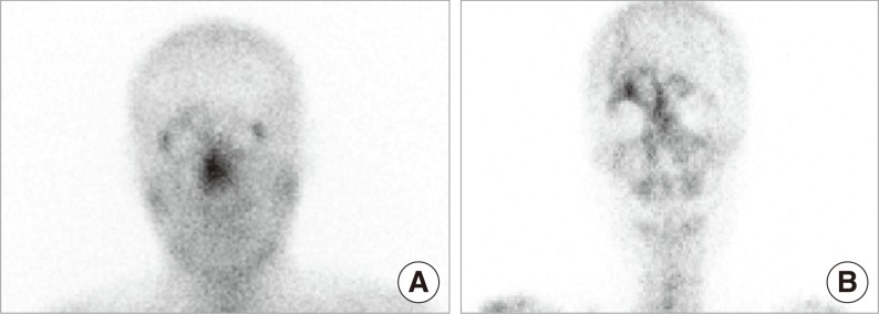
Fig. 4 Postoperative gallium scan (A) and technetium scan (B) one week after the surgery. The findings of both scans show increased uptake in the right frontal sinus area. However, uptake in the gallium scan was less increased than in the technetium scan. This finding was consistent with a resolved acute osteomyelitis.

Fig. 5 Same patient profile showing improvement of primary lesion without any defect six months after the operation.
Reference
-
1. Thomas JN, Nel JR. Acute spreading osteomyelitis of the skull complicating frontal sinusitis. J Laryngol Otol. 1977; 1. 91(1):55–62. PMID: 833490.2. Feder HM Jr, Cates KL, Cementina AM. Pott puffy tumor: a serious occult infection. Pediatrics. 1987; 4. 79(4):625–629. PMID: 3822683.3. Shehu BB, Mahmud MR. Pott's puffy tumour: a case report. Ann Afr Med. 2008; 9. 7(3):138–140. PMID: 19253524.
Article4. Maheshwar AA, Harris DA, Al-Mokhthar N, Evans RA. Pott's puffy tumour: an unusual presentation and management. J Laryngol Otol. 2001; 6. 115(6):497–499. PMID: 11429078.
Article5. Tattersall R, Tattersall R. Pott's puffy tumour. Lancet. 2002; 3. 23. 359(9311):1060–1063. PMID: 11937203.
Article6. Verbon A, Husni RN, Gordon SM, Lavertu P, Keys TF. Pott's puffy tumor due to Haemophilus influenzae: case report and review. Clin Infect Dis. 1996; 12. 23(6):1305–1307. PMID: 8953076.7. Deutsch E, Hevron I, Eilon A. Pott's puffy tumor treated by endoscopic frontal sinusotomy. Rhinology. 2000; 12. 38(4):177–180. PMID: 11190752.
- Full Text Links
-
- Actions
-
Cited
- CITED
-
- Close
- Share
-
- Similar articles
-
- A Case of External Approach Treatment of Pott's Puffy Tumor
- A Case of Pott's Puffy Tumor from Recurrent Upper Eyelid Abscess
- A case of Pott's puffy tumor with epidural abscess
- A Case of Pott's Puffy Tumor as a Complication of Contralateral Frontal Sinusitis
- A Case of Chronic Invasive Rhinocerebral Mucormycosis with Pott’s Puffy Tumor
