A Case of Huge Pulmonary Blastoma With Multiorgan Invasion
- Affiliations
-
- 1Department of Internal Medicine and Airway Remodeling Laboratory, Chonbuk National University Medical School, Jeonju, Republic of Korea. leeyc@chonbuk.ac.kr
- KMID: 2114604
- DOI: http://doi.org/10.4046/trd.2007.62.2.149
Abstract
- A pulmonary blastoma is a rare malignant tumor of the lung that is composed of epithelial and mesenchymal elements and resembles the structure of an embryonic lung. Pulmonary blastomas have a very poor prognosis and make up 0.25 to 0.5 percent of all primary malignant lung tumors. A pulmonary blastoma usually manifests as a solitary parenchymal mass or nodule and multiple subpleural mass with effusion on chest X-ray and computed tomography. We encountered a very rare case of pulmonary blastoma in a 52 years old male. He complained of abdominal pain, fullness, and dyspnea. The radiology examination revealed a huge lung mass invading the mediastinum, heart, diaphragm, and liver. The percutaneous needle biopsies were performed, and this tumor was diagnosed as a pulmonary blastoma. We report a biopsy confirmed case of a huge pulmonary blastoma invading multiple organs.
Keyword
MeSH Terms
Figure
-
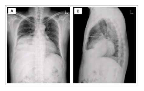
Figure 1 Chest radiographs show a huge mass on right lower lung field. (A) Posteroanterior view, (B) Lateral view of left side
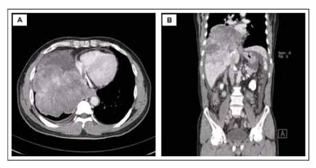
Figure 2 A CT scan shows a huge mass (14 × 12 × 11 cm) in the right lower lobe invading diaphragm and liver. (A) Enhanced axial view of CT scan (B) Coronal view of CT scan
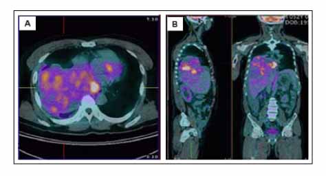
Figure 3 PET-CT scan shows unevenly hypermetabolic (p-SUV=8.34) huge mass in the right lower lobe invading mediastinum, diaphragm, and liver.
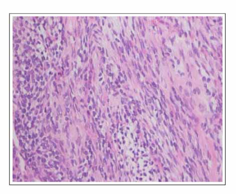
Figure 4 Photomicrograph of tissues obtained from percutaneous liver biopsy shows characteristic features of malignant stromal structure composed with undifferenciated small round cells and loose spindle cells. H&E stain (× 100).

Figure 5 Immunohistochemical stain for vimentin and CD99 of tissues obtained from percutaneous liver biopsy shows positive reaction in tumor cells (× 100).

Figure 6 Photomicrograph of tissues obtained from percutaneous transthoracic needle biopsy shows a malignant stromal structure composed with undifferenciated and immature celluar component. (A) H&E stain (× 100), (B) H&E stain (× 200).
Reference
-
1. Francis D, Jacobsen M. Pulmonary blastoma. Curr Top Pathol. 1983. 73:265–294.2. Barnett N, Barnard WG. Some unusual thoracic tumors. Br J Surg. 1945. 32:447–457.3. Liman ST, Altinok T, Topcu S, Tastepe AI, Uzarc A, Demircan S, et al. Survival of biphasic pulmonary blastoma. Respir Med. 2006. 100:1174–1179.4. LeMense GP, Reed CE, Silvestri GA. Pulmonary blastoma: a rare lung malignancy. Lung Cancer. 1996. 15:233–237.5. Di Lieto E, Baldi A, Vicidomini G, Di Marino MP, Baldi F. Pulmonary blastoma in adults. Minerva Chir. 1997. 52:839–846.6. Dogan R, Gungen Y, Ucanok K, Cetin G. Pulmonary blastoma. Hacettepe Med J. 1989. 22:235–239.7. Koss MN, Hochholzer L, O'Leary T. Pulmonary blastomas. Cancer. 1991. 67:2368–2381.8. Jacobsen M, Francis D. Pulmonary blastoma: aclinicopathological study of eleven cases. Acta Pathol Microbiol Scand. 1980. 88:151–160.9. Novotny JE, Hurias CM. Resection and adjuvant chemotherapy of pulmonary blastoma: a case report. Cancer. 1995. 76:1537–1539.10. Travis WD, Brambilla E, Muller-Hermelink HK, Harris CC. Small cell carcinoma, and sarcomatoid carcinoma. World Health Organization Classification of Tumours. Pathology and genetics of tumours of the lung, pleura, thymus and heart. 2004. Lyon: IARC Press;31–34. 53–58.11. Dienemann D, Hartmann CA, Minck C. Pulmonary blastomas: immunohistochemical investigations of three cases. Pathol Res Pract. 1989. 184:306–311.12. Adluri RK, Boddu SR, Martin-Ucar A, Duffy JP, Beggs FD, Morgan WE. Pulmonary blastoma: a rare tumor with variable presentation. Eur J Cardiothoracic Surg. 2006. 29:236–239.13. Zavala-Alarcou E, Sudhakar S, Gonzoles LR, Patel R. Extension of a pulmonary blastoma into the left atrium. Mayo Clin Proc. 2001. 76:657–660.14. Lee HJ, Goo JM, Kim KW, Im JG, Kim JH. Pulmonary blastoma:radiologic findings in five patients. Clin Imaging. 2004. 28:113–118.15. Robert J, Pache JC, Seium Y, de Perrot M, Spiliopoulos A. Pulmonary blastoma: report of five cases and identification of clinical features suggestive of the disease. Eur J Cardiothorac Surg. 2002. 22:708–711.
