Upfront Stereotactic Radiosurgery for Pineal Parenchymal Tumors in Adults
- Affiliations
-
- 1Department of Neurosurgery, Asan Medical Center, University of Ulsan College of Medicine, Seoul, Korea. yhyunc@amc.seoul.kr
- 2Department of Pathology, Asan Medical Center, University of Ulsan College of Medicine, Seoul, Korea.
- KMID: 2114369
- DOI: http://doi.org/10.3340/jkns.2015.58.4.334
Abstract
OBJECTIVE
Pineal parenchymal tumors (PPTs) in adults are rare, and knowledge regarding their optimal management and treatment outcome is limited. Herein, we present the clinical results of our series of PPTs other than pineoblastomas managed by stereotactic radiosurgery (SRS) at upfront setting.
METHODS
Between 1997 and 2014, nine consecutive adult patients with the diagnosis of PPTs, either pineocytoma or pineal parenchymal tumor of intermediate differentiation, were treated with SRS. There were 6 men and 3 women. The median age was 39 years (range, 31-53 years). All of the patients presented with symptoms of hydrocephalus. Endoscopic third ventriculostomy and biopsy was done for initial management. After histologic diagnosis, patients were treated with Gamma Knife with the mean dose of 13.3 Gy (n=3) or fractionated Cyberknife with 32 Gy (n=6).
RESULTS
After a mean follow-up of 78.6 months (range, 14-223 months), all patients were alive and all of their tumors were locally controlled except for one instance of cerebrospinal fluid seeding metastasis. On magnetic resonance images, tumor size decreased in all patients, resulting in complete response in 3 patients and partial response in 6. One patient had experienced temporary memory impairment after SRS, which improved spontaneously.
CONCLUSION
SRS is effective and safe for PPTs in adults and can be considered as a useful alternative to surgical resection at upfront setting.
Keyword
MeSH Terms
Figure
-
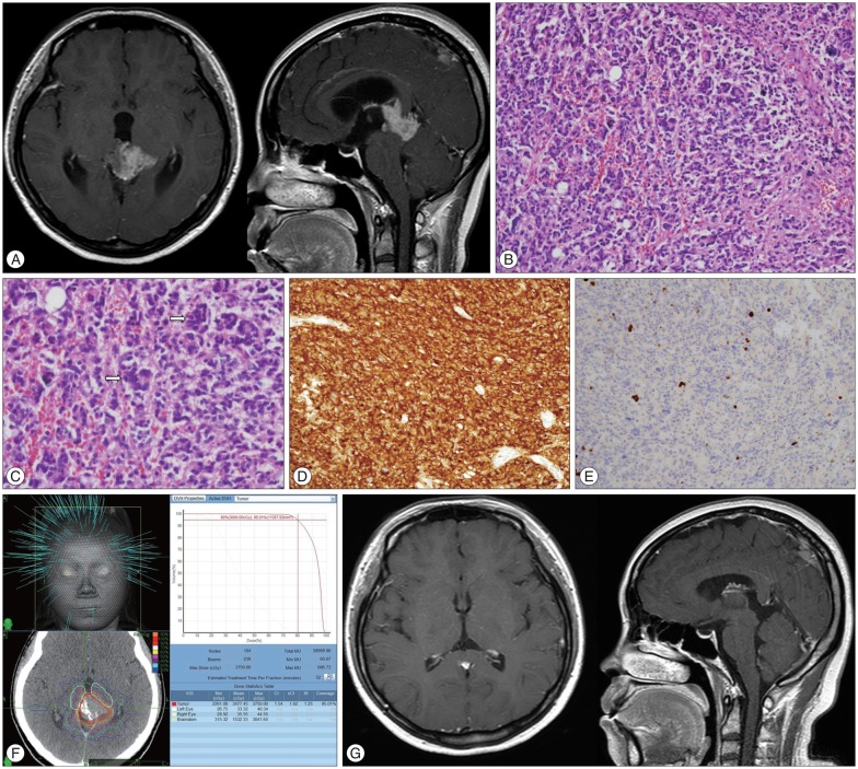
Fig. 1 A 34-year-old woman with pineal parenchymal tumor of intermediate differentiation (Case No. 7). A : Gadolinium-enhanced magnetic resonance images showing a 3.2×2.4×2.0 cm sized tumor in the pineal region with obstructive hydrocephalus. B : Tumor shows diffusely high cellularity and tumor cell nuclei are pleomorphic. C : Multinucleated giant tumor cells are frequently seen (arrows). D : Tumor cells are diffusely immunoreactive for synaptophysin. E : Proliferation activity assessed by MIB-1 is low. F : She received 5-fraction Cyberknife (CK) treatment with marginal dose of 30 Gy. G : Tumor disappeared completely in 9 months after CK, and no evidence of disease was seen at the last follow-up of 3 years.
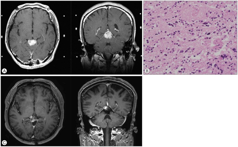
Fig. 2 A 44-year-old man with pineocytoma (Case No. 2). A : Gadolinium-enhanced magnetic resonance images at the time of Gamma Knife (GK) treatment. B : Tumor consists of relatively small, uniform, mature cells resembling normal pineocytes, and cell-free spaces filled with cell processes are forming vague rosettes. C : After GK treatment with marginal dose of 12 Gy, he achieved durable tumor response with more than 18 years of follow-up.
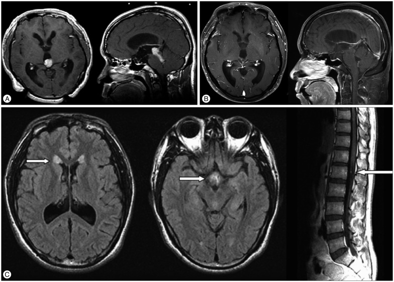
Fig. 3 A 31-year-old man with pineal parenchymal tumor of intermediate differentiation (Case No. 1). Gadolinium-enhanced magnetic resonance images before (A) and 84 months after Gamma Knife treatment (B). Although the primary site remained free of disease at this time, cerebrospinal fluid seeding metastases were observed in the ventricles and the cauda equina (C, arrows). He received additional craniospinal irradiation and his disease was stable at 17 years.
Reference
-
1. Al-Hussaini M, Sultan I, Abuirmileh N, Jaradat I, Qaddoumi I. Pineal gland tumors : experience from the SEER database. J Neurooncol. 2009; 94:351–358. PMID: 19373436.
Article2. Anan M, Ishii K, Nakamura T, Yamashita M, Katayama S, Sainoo M, et al. Postoperative adjuvant treatment for pineal parenchymal tumour of intermediate differentiation. J Clin Neurosci. 2006; 13:965–968. PMID: 16904896.
Article3. Blakeley JO, Grossman SA. Management of pineal region tumors. Curr Treat Options Oncol. 2006; 7:505–516.
Article4. Bruce JN, Ogden AT. Surgical strategies for treating patients with pineal region tumors. J Neurooncol. 2004; 69:221–236. PMID: 15527093.
Article5. Bruce JN, Stein BM. Surgical management of pineal region tumors. Acta Neurochir (Wien). 1995; 134:130–135. PMID: 8748771.
Article6. Chandy MJ, Damaraju SC. Benign tumours of the pineal region : a prospective study from 1983 to 1997. Br J Neurosurg. 1998; 12:228–233. PMID: 11013685.7. Clark AJ, Ivan ME, Sughrue ME, Yang I, Aranda D, Han SJ, et al. Tumor control after surgery and radiotherapy for pineocytoma. J Neurosurg. 2010; 113:319–324. PMID: 20136388.
Article8. Clark AJ, Sughrue ME, Aranda D, Parsa AT. Contemporary management of pineocytoma. Neurosurg Clin N Am. 2011; 22:403–407. PMID: 21801989.
Article9. Fauchon F, Jouvet A, Paquis P, Saint-Pierre G, Mottolese C, Ben Hassel M, et al. Parenchymal pineal tumors : a clinicopathological study of 76 cases. Int J Radiat Oncol Biol Phys. 2000; 46:959–968. PMID: 10705018.10. Fontana EJ, Garvin J, Feldstein N, Anderson RC. Pediatric considerations for pineal tumor management. Neurosurg Clin N Am. 2011; 22:395–402. PMID: 21801988.
Article11. Han SJ, Clark AJ, Ivan ME, Parsa AT, Perry A. Pathology of pineal parenchymal tumors. Neurosurg Clin N Am. 2011; 22:335–340. PMID: 21801981.
Article12. Hasegawa T, Kondziolka D, Hadjipanayis CG, Flickinger JC, Lunsford LD. The role of radiosurgery for the treatment of pineal parenchymal tumors. Neurosurgery. 2002; 51:880–889. PMID: 12234394.
Article13. Hernesniemi J, Romani R, Albayrak BS, Lehto H, Dashti R, Ramsey C 3rd, et al. Microsurgical management of pineal region lesions : personal experience with 119 patients. Surg Neurol. 2008; 70:576–583. PMID: 19055952.
Article14. Ito T, Kanno H, Sato K, Oikawa M, Ozaki Y, Nakamura H, et al. Clinicopathologic study of pineal parenchymal tumors of intermediate differentiation. World Neurosurg. 2014; 81:783–789. PMID: 23396072.
Article15. Kang JK, Jeun SS, Hong YK, Park CK, Son BC, Lee IW, et al. Experience with pineal region tumors. Childs Nerv Syst. 1998; 14:63–68. PMID: 9548344.
Article16. Kano H, Niranjan A, Kondziolka D, Flickinger JC, Lunsford D. Role of stereotactic radiosurgery in the management of pineal parenchymal tumors. Prog Neurol Surg. 2009; 23:44–58. PMID: 19329860.
Article17. Komakula S, Warmuth-Metz M, Hildenbrand P, Loevner L, Hewlett R, Salzman K, et al. Pineal parenchymal tumor of intermediate differentiation : imaging spectrum of an unusual tumor in 11 cases. Neuroradiology. 2011; 53:577–584. PMID: 21080159.
Article18. Konovalov AN, Pitskhelauri DI. Principles of treatment of the pineal region tumors. Surg Neurol. 2003; 59:250–268. PMID: 12748006.
Article19. Lapras C, Patet JD, Mottolese C, Lapras C Jr. Direct surgery for pineal tumors : occipital-transtentorial approach. Prog Exp Tumor Res. 1987; 30:268–280. PMID: 3628812.20. Lekovic GP, Gonzalez LF, Shetter AG, Porter RW, Smith KA, Brachman D, et al. Role of Gamma Knife surgery in the management of pineal region tumors. Neurosurg Focus. 2007; 23:E12. PMID: 18081477.
Article21. Louis DN, Ohgaki H, Wiestler OD, Cavenee WK, Burger PC, Jouvet A, et al. The 2007 WHO classification of tumours of the central nervous system. Acta Neuropathol. 2007; 114:97–109. PMID: 17618441.
Article22. Luo SQ, Li DZ, Zhang MZ, Wang ZC. Occipital transtentorial approach for removal of pineal region tumors : report of 64 consecutive cases. Surg Neurol. 1989; 32:36–39. PMID: 2734686.
Article23. Lutterbach J, Fauchon F, Schild SE, Chang SM, Pagenstecher A, Volk B, et al. Malignant pineal parenchymal tumors in adult patients : patterns of care and prognostic factors. Neurosurgery. 2002; 51:44–55. discussion 55-56PMID: 12182434.24. Macdonald DR, Cascino TL, Schold SC Jr, Cairncross JG. Response criteria for phase II studies of supratentorial malignant glioma. J Clin Oncol. 1990; 8:1277–1280. PMID: 2358840.
Article25. Mori Y, Kobayashi T, Hasegawa T, Yoshida K, Kida Y. Stereotactic radiosurgery for pineal and related tumors. Prog Neurol Surg. 2009; 23:106–118. PMID: 19329865.
Article26. Reyns N, Hayashi M, Chinot O, Manera L, Péragut JC, Blond S, et al. The role of Gamma Knife radiosurgery in the treatment of pineal parenchymal tumours. Acta Neurochir (Wien). 2006; 148:5–11. discussion 11PMID: 16172830.
Article27. Sato K, Kubota T. Pathology of pineal parenchymal tumors. Prog Neurol Surg. 2009; 23:12–25. PMID: 19329858.
Article28. Selvanathan SK, Hammouche S, Smethurst W, Salminen HJ, Jenkinson MD. Outcome and prognostic features in adult pineoblastomas : analysis of cases from the SEER database. Acta Neurochir (Wien). 2012; 154:863–869. PMID: 22460262.
Article29. Vaquero J, Ramiro J, Martínez R, Bravo G. Neurosurgical experience with tumours of the pineal region at Clinica Puerta de Hierro. Acta Neurochir (Wien). 1992; 116:23–32. PMID: 1319669.
Article
