J Korean Soc Magn Reson Med.
2012 Dec;16(3):266-270. 10.13104/jksmrm.2012.16.3.266.
Small Cell Carcinoma of the Gallbladder: A Case Report
- Affiliations
-
- 1Department of Radiology, Wonkwang University School of Medicine and Hospital, Korea. yjyh@wonkwang.ac.kr
- 2Department of Pathology, Wonkwang University School of Medicine and Hospital, Korea.
- KMID: 2099864
- DOI: http://doi.org/10.13104/jksmrm.2012.16.3.266
Abstract
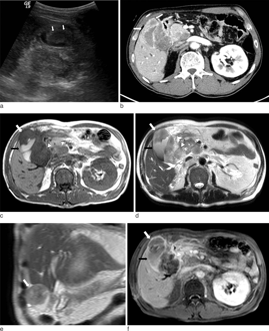
- Small cell carcinoma of the gallbladder is a type of neuroendocrine tumor and very rare. We report ultrasound, CT and MR findings of a small cell carcinoma of the gallbladder that was confirmed by pathology. Small cell carcinoma of the gallbladder was seen as a well-defined mass with peripheral rim enhancement in the gallbladder. In spite of the large size of the mass, direct and extensive invasion of the liver was not detected. However, there were many metastatic lymph nodes.
Keyword
Figure
Reference
-
1. Lee MH, Park TJ, Lee HW, et al. Small cell carcinoma of the gall bladder. Two case reports. Korean J Med. 2007. 73:1022–1028.2. Albores-Saavedra J, Cruz-Ortis H, Alcantra-Vazques A, Henson DE. Unusal types of gallbladder carcinoma. A report of 16 cases. Arch Pathol Lab Med. 1981. 105:287–293.3. Kuwabara H, Uda H. Small cell carcinoma of the gall-bladder with intestinal metaplastic epithelium. Pathol Int. 1998. 48:303–306.4. Moskal TL, Zhang PJ, Nava HR. Small cell carcinoma of the gallbladder. J Surg Oncol. 1999. 70:54–59.5. Cavazzana AO, Fassina AS, Tollot M, Ninfo V. Small-cell carcinoma of gallbladder. An immunocytochemical and ultrasrtuctural study. Pathol Res Pract. 1991. 187:472–476.6. Choi WB, Lee TY, Lee NW, et al. A case of small cell carcinoma of gallbladder. Korean J Med. 1997. 53:847–852.7. Obuz F, Altay C, Sagol O, Astarcioglu H, Oztop I, Igci E. MDCT findings in neuroendocrine carcinoma of the gallbladder: case report. Abdom Imaging. 2007. 32:105–107.8. Ahn JE, Byun JH, Ko MS, Park SH, Lee MG. Neuroendocrine carcinoma of the gallbladder causing hyperinsulinaemic hypoglycaemia. Clinical Radiology. 2007. 62:391–394.


