Fibrolipomatous Hamartoma of Digital Branch of the Median Nerve without Macrodystrophy: Two Case Reports with Magnetic Resonance Imaging Findings
- Affiliations
-
- 1Department of Radiology, Chung-Ang University Hospital, School of Medicine, Chung-Ang University, Seoul, Korea. jyj907075@naver.com
- 2Department of Orthopedic Surgery, Chung-Ang University Hospital, School of Medicine, Chung-Ang University, Seoul, Korea.
- 3Department of Pathology, Chung-Ang University Hospital, School of Medicine, Chung-Ang University, Seoul, Korea.
- KMID: 2097985
- DOI: http://doi.org/10.3348/jksr.2012.67.6.483
Abstract
- Fibrolipomatous hamartoma (FLH) of the nerve is a rare, benign tumor that most commonly originates from the median nerve of the hand. Fibrofatty tissue proliferates around the nerve and infiltrates the epineurium and perineurium. We present two cases of pathologically proven FLH of a digital branch of the median nerve, without macrodystrophy with magnetic resonance imaging, surgical and pathologic findings. Magnetic resonance images of both cases show well-circumscribed mass with fat signal intensity around an enlarged digital branch of the median nerve and characteristic coaxial-cable-like appearance on axial images and spaghetti-like appearance on coronal images.
MeSH Terms
Figure
-
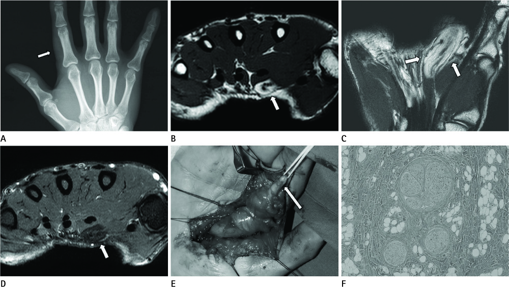
Fig. 1 A 39-year-old man with pathologically proven fibrolipomatous hamartoma involving digital branch of median nerve at right second finger. Radiograph (A) shows soft tissue bulging in radial aspect of right second finger (arrow) without bony abnormality. Axial T1-weighted (B) and coronal T2-weighted images (C) show enlargement of the digital branch of the median nerve and hypointense nerve bundles embedded within fat (arrows). Axial fat-suppressed T1 weighted image with gadolinium enhancement (D) shows non-enhancing hypointense fat tissue (arrow) around the digital branch of the median nerve. Intraoperative photograph (E) shows fusiform shaped mass and digital nerve fiber (arrow) at the distal ends of the mass. Photomicrograph (F) shows fibrofatty tissue growth surrounding the nerve trunk. Septation of nerve fascicles and concentric perineural fibrosis are noted (Hematoxylin & Eosin staining, × 100).
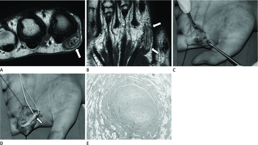
Fig. 2 A 31-year-old man with pathologically proven fibrolipomatous hamartoma involving digital branch of median nerve at left second finger. Axial T2-weighted images (A) shows marked enlargement of the digital branch of the median nerve (arrow) surrounded by prominent fat tissue. Coronal T2-weighted images (B) shows hypointense nerve bundles embedded within fat (arrows). Intraoperative photograph (C) shows a yellowish fusiform shaped mass involving digital branch of median nerve at left second finger. Another intraoperative photograph (D) shows that the continuity of the digital nerve (arrow) was preserved after debulking of the fibrolipomatous hamartoma of the nerve. Photomicrograph (E) shows adipose and fibrous tissue surrounds and infiltrates the nerve trunk, causing enlargement of the nerve (Hematoxylin & Eosin staining, × 40).
Reference
-
1. Silverman TA, Enzinger FM. Fibrolipomatous hamartoma of nerve. A clinicopathologic analysis of 26 cases. Am J Surg Pathol. 1985. 9:7–14.2. De Maeseneer M, Jaovisidha S, Lenchik L, Witte D, Schweitzer ME, Sartoris DJ, et al. Fibrolipomatous hamartoma: MR imaging findings. Skeletal Radiol. 1997. 26:155–160.3. Marom EM, Helms CA. Fibrolipomatous hamartoma: pathognomonic on MR imaging. Skeletal Radiol. 1999. 28:260–264.4. Kransdorf MJ. Benign soft-tissue tumors in a large referral population: distribution of specific diagnoses by age, sex, and location. AJR Am J Roentgenol. 1995. 164:395–402.5. Toms AP, Anastakis D, Bleakney RR, Marshall TJ. Lipofibromatous hamartoma of the upper extremity: a review of the radiologic findings for 15 patients. AJR Am J Roentgenol. 2006. 186:805–811.6. Terzis JK, Daniel RK, Williams HB, Spencer PS. Benign fatty tumors of the peripheral nerves. Ann Plast Surg. 1978. 1:193–216.7. Cui Q, Chhabra AB, Leo BM, Pannunzio ME. Lipofibromatous hamartoma of a digital nerve. Am J Orthop (Belle Mead NJ). 2008. 37:E146–E148.8. Boren WL, Henry RE Jr, Wintch K. MR diagnosis of fibrolipomatous hamartoma of nerve: association with nerve territory-oriented macrodactyly (macrodystrophia lipomatosa). Skeletal Radiol. 1995. 24:296–297.9. Zeng R, Frederick-Dyer K, Ferguson NL, Lewis J, Fu Y. Fibrolipomatous hamartoma of the inferior calcaneal nerve (Baxter nerve). Skeletal Radiol. 2012. 41:1323–1326.10. Murphey MD, Smith WS, Smith SE, Kransdorf MJ, Temple HT. From the archives of the AFIP. Imaging of musculoskeletal neurogenic tumors: radiologic-pathologic correlation. Radiographics. 1999. 19:1253–1280.11. Gundes H, Alici T, Sahin M. Neural fibrolipoma of the digital nerve: a case report. J Orthop Surg (Hong Kong). 2011. 19:123–125.
- Full Text Links
-
- Actions
-
Cited
- CITED
-
- Close
- Share
-
- Similar articles
-
- Fibrolipomatous hamartoma of the median nerve: A case report
- Intradural Spinal Fibrolipomatous Hamartoma: A Case Report
- The Change of Preoperative and Postoperaive Magnetic Resonance Imaging Findings in Lipofibromatous Hamartoma of the Median Nerve
- Invasive Ductal Carcinoma Arising within a Mammary Hamartoma: Case Report
- Symmetric Lipofibromatous Hamartoma Affecting Digital Nerves
