Radiological Features of Soft Tissue Calcification: A Pictorial Essay
- Affiliations
-
- 1Department of Radiology, Daejeon St. Mary's Hospital, The Catholic University of Korea, Korea. yslee1074@catholic.ac.kr
- KMID: 2097906
- DOI: http://doi.org/10.3348/jksr.2010.63.3.275
Abstract
- Calcification of soft tissue may represent a nonspecific local response or be a manifestation of underlying disease. The diagnosis and principle of treatment varies depending on the characteristics of the calcified lesions. Soft tissue calcification is classified by mechanism into 3 types: metastatic calcifications, calcinosis, and dystrophic calcifications. However, classification according to the site and shape of calcification may be more helpful for a clinical diagnosis. The purpose of this pictorial essay is to classify soft tissue calcifications according to their location: vessel, periarticular, joint capsule, tendon, bursa, cartilage, ligament, and in tumors, as well as to document the characteristic radiological findings and causes of calcifications.
MeSH Terms
Figure
-
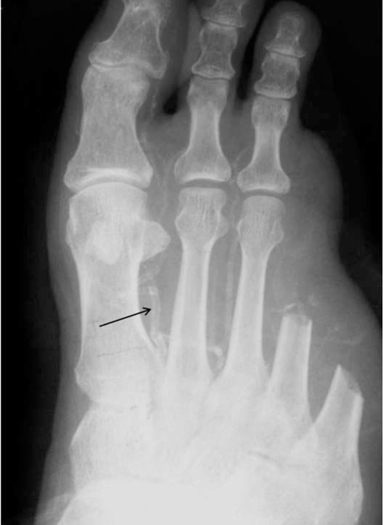
Fig. 1 DM foot in a 63-year-old man with skin necrosis. Anteroposterior (AP) plain radiograph of foot shows tubular, elongated arterial calcifications (arrow).
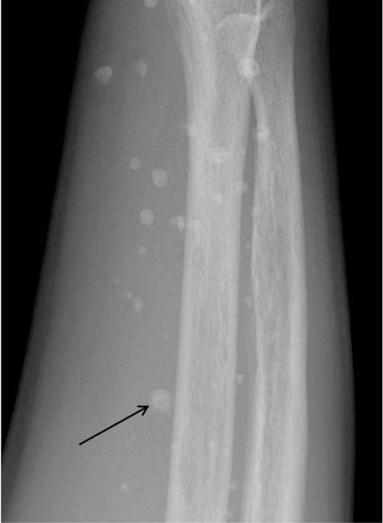
Fig. 2 Cavernous hemangioma in a 20-year-old man. Lateral plain radiograph of forearm shows several round, ring like calcifications including central lucencies (arrow), suggesting thromboliths.

Fig. 3 Calcifications with venous stasis in a 46-year-old man. AP plain radiograph of tibia reveals stripe like shadows of calcifications (arrow).
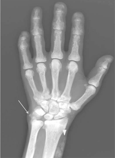
Fig. 4 Renal osteodystrophy in a 48-year-old woman. Posteroanterior (PA) plain radiograph of hand shows several nodular calcifications along the periarticular, periosteal spaces and subperiosteal resorptions in the phalangeal bones. Globular calcifications are also seen in the ulnocarpal periarticular area (arrow) and soft tissue (arrowhead) of the distal radial level.

Fig. 5 Pseudohypoparathyroidism in an 11-year-old female with delayed development, mental retardation and short stature. Plain radiograph of hand shows a periarticular calcification adjacent to fourth metacarpophalangeal joint (arrow). Positive metacarpal sign is also seen (line).

Fig. 6 Calcific tendinitis in a 53-yearold man in the shoulder. A. AP plain radiograph of right shoulder shows a globular calcified density (arrow) adjacent to the greater tuberosity of humerus. B. Coronal fat suppressed T1-weighted MR arthrography reveals a dark signal intensity lesion (arrow) in the distal supraspinatus tendon, suggesting calcific tendinitis.
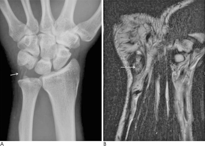
Fig. 7 Calcific tendinitis of the flexor carpi ulnaris tendon in a 36-year-old man. A. Plain radiograph of wrist show irregular, linear calcified densities (arrow) in the ulnocarpal space. B. Coronal T2-weighted MR image of wrist shows high signal intensity including dark signal (arrow) at the pisiform insertion portion of flexor carpi ulnaris tendon, suggesting calcific tendinitis.

Fig. 8 Calcific tendinitis of right hip in a 56-year-old woman. A. Plain radiograph of right hip shows a globular appearance of calcified density (arrow) adjacent to the greater trochanter. B, C. Coronal (B) and axial (C) fat suppressed T2-weighted MR images of hip reveal the calcification (arrow) in the femoral insertion portion of gluteus medius tendon as well as the high signal intensity surrounding dark signal, which is consistent with inflammation.
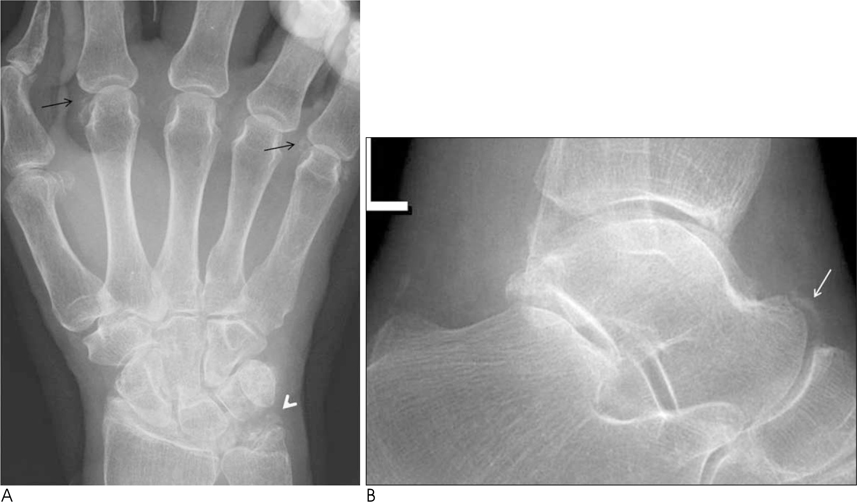
Fig. 9 CPPD in an 83-year-old woman. A. Plain radiograph of hand shows nodular calcifications (arrows) along the MCP joints and triangular calcifications (arrowhead) in the ulnocarpal joint, representing TFCC calcifications. B. Lateral radiograph of ankle shows a curvilinear calcification (arrow) along the talonavicular joint.
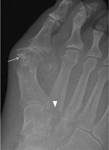
Fig. 10 Gouty arthritis in a 42-year-old man. Plain radiograph of foot reveals bony erosion including stippled calcifications (arrow) at the first medial metatarsal head. Another well defined bony erosion with small calcifications (arrowhead) in the second metatarso-cuneiform joint.

Fig. 11 CREST syndrome in a 62-year-old woman. A. Lateral radiograph of forearm shows a nodular and lobulated calcification (arrow) in the subcutaneous tissue. B. Lateral radiograph of knee reveals stippled calcifications (arrow) in the subcutaneous tissue of prepatellar region. C. Sagittal T2-weighted MR image of knee shows several dark signals (arrow) in the prepatellar subcutaneous area.
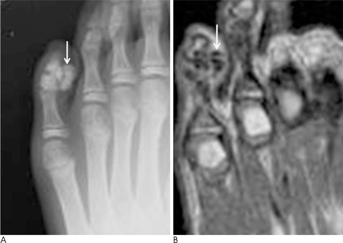
Fig. 12 Tumoral calcinosis in a 12-year-old man. A. AP plain radiograph of foot shows multiloculated calcified juxta-articular mass (arrow) adjacent to DIP joint of little toe. B. Axial T1-weighted MR image of foot shows several calcified nodules with dark signal intensity (arrow) around the DIP joint of little toe.
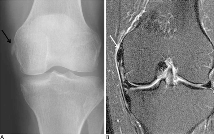
Fig. 13 Pellegrini Stieda disease in a 58-year-old woman. A. Plain radiograph of knee reveals an ovoid shaped calcification (arrow) adjacent to medial femoral condyle. B. Coronal fat suppressed T2-weighted MR image of knee shows a nodular dark signal intensity lesion (arrow) in the thickened medial collateral ligament (MCL) at the femoral insertion portion, suggesting chronic injury of MCL with calcification.
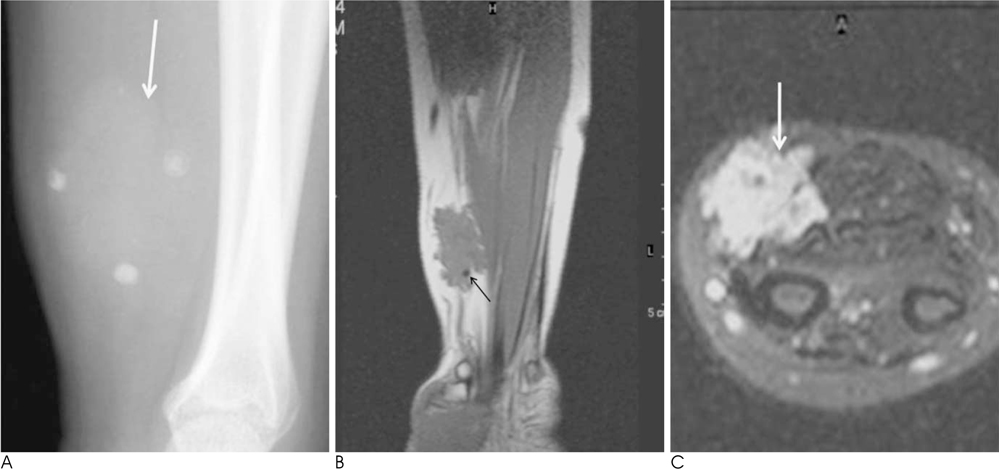
Fig. 14 Cavernous hemangioma in a 25-year-old woman. A. Plain radiograph of forearm shows a soft tissue mass including several phleboliths (arrow) in the volar aspect of forearm. B. Coronal T1-weighted MR image of forearm demonstrates a multilobular contour of the soft tissue mass and internal signal voids (arrow) due to phleboriths. C. Axial fat suppressed T2-weighted MR image reveals a soft tissue mass of multilobulated, multiseptated high signal intensity, containing several calcifications (arrow).

Fig. 15 Well differentiated liposarcoma in a 42-year-old man. A. Lateral radiograph of femur shows a soft tissue mass of the fatty nature including nonspecific calcifications (arrow) and ossifications in the posterior aspect of distal femur. B. Coronal T1-weighted MR image of thigh reveals a mainly high signal intensity mass with irregular septa and dark signals (arrows), suggesting calcifications. C. Coronal fat suppressed T2-weighted MR image shows fat suppression of the adipose tissue and increased signal intensity of no adipose area in the superomedial aspect of mass (arrow).
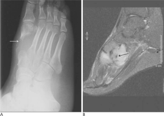
Fig. 16 Synovial sarcoma in a 40-year old man. A. Plain radiograph of foot shows bony destruction including irregular small calcifications (arrow) in the first metatarsal bone. B. Sagittal fat suppressed T2-weighted MR image demonstrates well defined, a lobulated mass in the medial midfoot including calcification (arrow). The signal intensity of mass shows heterogeneous signal intensity.
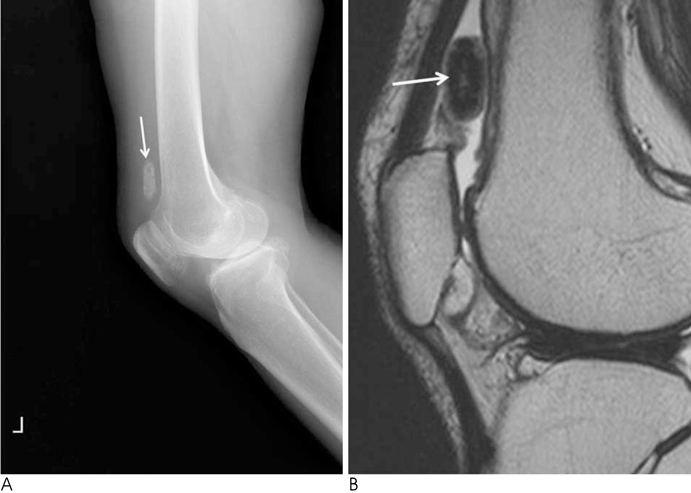
Fig. 17 Synovial chondroma in a 56-year-old woman. A. Plain lateral radiograph of knee shows an ovoid shaped calcified nodule (arrow) in the suprapatellar area. B. Sagittal T2-weighted MR image reveals an ovoid mass dark signal intensity including central high signal intensity (arrow) in the fluid filled suprapatellar bursa.
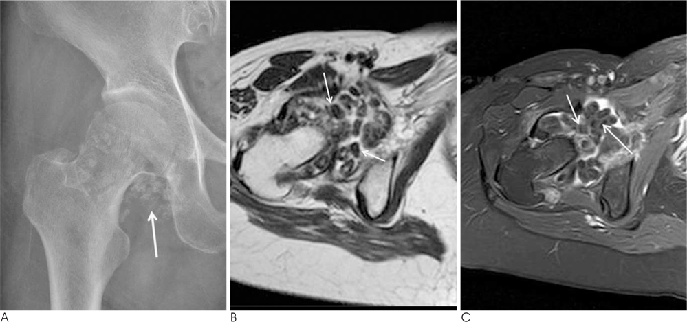
Fig. 18 Synovial chondromatosis in a 62-year-old woman. A. Plain radiograph shows multiple calcifications of right hip (arrow). B. Axial T2-weighted MR image of hip reveals multiple loose bodies (arrows). C. Axial enhanced fat suppressed T1-weighted MR image demonstrates many loose bodies (arrows) without contrast enhancement in the enhanced synovial fluid.
Reference
-
1. Gartner L, Pearce CJ, Saifuddin A. The role of the plain radiograph in the characterisation of soft tissue tumours. Skeletal Radiol. 2009; 38:549–558.2. Resnick D. Diagnosis of bone and joint disorders. 4th ed. Philadelphia: Saunders;2002. p. 4635–4665.3. Hussmann J, Russell RC, Kucan JO, Khardori R, Steinau HU. Soft tissue calcifications: differential diagnosis and therapeutic approaches. Ann Plast Sur. 1995; 34:138–147.4. Boulman N, Slobodin G, Rozenbaum M, Rosner I. Calcinosis in rheumatic disease. Semin Arthritis Rheum. 2005; 34:805–812.5. Jevtic V. Imaging of renal osteodystrophy. Eur J Radiol. 2003; 46:85–95.6. Jung CB, Gentili A, Chew FS. Calcific tendinosis and periarthritis. Classic magnetic resonance imaging appearance and associated findings. J Comput Assit Tomogr. 2004; 28:390–396.7. Saffar P. Chondrocalcinosis of the wrist. J Hand Surg Br. 2004; 29:486–493.8. Resnick D. Calcium Pyrophosphate Dihydrate Crystal Deposition Disease. In : Resnick D, editor. Diagnosis of bone and joint disorders. 4th ed. Philadelphia: Saunders;2002. p. 1564–1565.9. Smack D, Norton SA, Fitzpatrick JE. Proposal for a pathogenesis-based classification of tumoral calcinosis. Int J Dermatol. 1996; 36:265–271.10. Kransdorf MJ, Murphey MD. Imaging of soft tissue tumors. Philadelphia: W.B. Saunders Co.;1997. p. 103–118.11. Kransdorf MJ, Murphey MD. Imaging of soft tissue tumors. 2nd ed. Philadelphia: Lippincott Williams & Wilkins;2006. p. 80–149.12. Kind M, Stock N, Coindre JM. Histology and imaging of soft tissue sarcomas. Eur J Radiol. 2009; 72:6–15.13. Kransdorf MJ, Murphey MD. Imaging of soft tissue tumors. 2nd ed. Philadelphia: Lippincott Williams & Wilkins;2006. p. 457–460.
- Full Text Links
-
- Actions
-
Cited
- CITED
-
- Close
- Share
-
- Similar articles
-
- Imaging Features of Soft-Tissue Calcifications and Related Diseases: A Systematic Approach
- Imaging Features of the Mesenchymal Tumors of the Breast according to WHO Classification: A Pictorial Essay
- Unusual, but important, peri- and extra-articular manifestations of rheumatoid arthritis: a pictorial essay
- Breast lesions during pregnancy and lactation: a pictorial essay
- Multi-Detector CT Findings of Typical and Atypical Appendicitis: A Pictorial Essay
