Inflammatory Myofibroblastic Tumor of the Bladder: Report of Two Cases
- Affiliations
-
- 1Department of Radiology, The Catholic University of Korea, Korea. hiohsn@catholic.ac.kr
- 2Department of Hospital Pathology, The Catholic University of Korea, Korea.
- KMID: 2097903
- DOI: http://doi.org/10.3348/jksr.2010.63.3.261
Abstract
- Inflammatory myofibroblastic tumor (IMT) is a rare condition of unknown origin. Pathologically, the lesion is composed of myofibroblastic spindle cells accompanied by an inflammatory infiltrate of plasma cells, lymphocytes, and eosinophils. We report two cases of inflammatory myofibroblastic tumor of the bladder which showed different imaging features and was falsely diagnosed as malignant tumors. We discuss the imaging findings along with a literature review.
Figure
-

Fig. 1 A 28-year-old woman presented with inflammatory myofibroblastic tumor. Color Doppler sonogram (A) shows a hypoechoic and hypervascular mass in the anterior wall of the bladder dome. Contrast-enhanced axial (B) and sagittal (C) CT scans show a well contrast enhancing portion (long arrow in B and C) on the right side of the mass. Moreover, a poorly enhancing area (asterisk) was evident in the left posterior portion. An irregular-shaped lesion (open arrow in B) was seen in the dependent portion of the contrast-filled bladder, suggesting hematoma. Microphotography (D) shows a proliferation of atypical spindle cells (arrows) and inflammatory cell infiltration in a myxoid background.
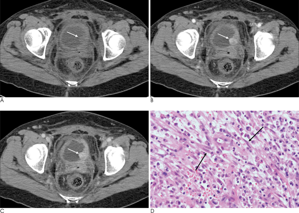
Fig. 2 A 51-year-old woman with an inflammatory myofibroblastic tumor. Dynamic contrast-enhanced axial CT scans show a rounded mass (arrows in A-C) with strong enhancement on arterial (B) and portal (C) phase, arising from the left lateral aspect of the bladder. Note the diffuse and irregular bladder wall thickening. Microphotography (D) shows a proliferation of atypical spindle cells (arrows) and inflammatory cell infiltration with a myxoid background.
Reference
-
1. Wong-You-Cheong JJ, Woodward PJ, Manning MA, Davis CJ. Inflammatory and non-neoplastic bladder masses: radiologicpathologic correlation. Radiographics. 2006; 26:1847–1868.2. Kumar A, Bhatti SS, Sharma S, Gupta SD, Kumar R. Inflammatory pseudotumor of urinary bladder: a diagnostic and management dilemma. Int Urol Nephrol. 2007; 39:799–802.3. Narla LD, Newman B, Spottswood SS, Narla S, Kolli R. Inflammatory pseudotumor. Radiographics. 2003; 23:719–729.4. Mochizuki Y, Kanda S, Nomata K, Hayashi T, Yamasaki Y, Kanetake H, et al. Spontaneous regression of inflammatory pseudotumor of the urinary bladder. Urol Int. 1999; 63:255–257.5. Kim SH, Cho JY, Kim SH. Inflammatory myofibroblastic pseudotumor of the urinary bladder in a patient with bilateral renal cell carcinoma. J Ultrasound Med. 2008; 27:483–486.6. Park SB, Cho KS, Kim JK, Lee JH, Jeoung AK, Kwon WJ, et al. Inflammatory pseudotumor (myoblastic tumor) of genitourinary tract. AJR Am J Roentgenol. 2008; 191:1255–1262.7. Houben CH, Chan A, Lee KH, Tam YH, To KF, Cheung W, et al. Inflammatory myofibroblastic tumor of the bladder in children: what can be expected. Pediatr Surg Int. 2007; 23:815–819.8. Sugita R, Saito M, Miura M, Yuda F. Inflammatory pseudotumour of the bladder: CT and MRI findings. Br J Radiol. 1999; 72:809–811.9. Young RH. Tumor-like lesions of the urinary bladder. Mod Pathol. 2009; 22:Suppl 2. S37–S52.10. Ymamoto H, Yamaguchi H, Aishima S, Oda Y, Kohashi K, Oshiro Y, et al. Inflammatory myofibroblastic tumor versus IgG4-related sclerosing disease and inflammatory pseudotumor: a comparative clinicopathologic study. Am J Surg Pathol. 2009; 33:1330–1340.
- Full Text Links
-
- Actions
-
Cited
- CITED
-
- Close
- Share
-
- Similar articles
-
- Inflammatory Myofibroblastic Tumor of Nasal Septum after Septoplasty: A Case Report
- Inflammatory Myofibroblastic Tumor of Kidney
- Inflammatory Myofibroblastic Tumor of the Urinary Bladder in a Young Patient With Gross Hematuria: A Case Report
- A Case of Cutaneous Inflammatory Myofibroblastic Tumor
- A Case of Inflammatory Myofibroblastic Tumor of the Urethra with Overactive Bladder
