Mucosal dehiscence coverage for dental implant using sprit pouch technique: a two-stage approach
- Affiliations
-
- 1Second Department of Comprehensive Care, Tsurumi University School of Dental Medicine, Yokohama, Japan.
- 2Unit of Oral and Maxillofacial Implantology, Tsurumi University School of Dental Medicine, Yokohama, Japan. ueno-d@tsurumi-u.ac.jp
- KMID: 2094734
- DOI: http://doi.org/10.5051/jpis.2012.42.3.105
Abstract
- PURPOSE
Soft tissue recessions frequently cause esthetic disharmony and dissatisfaction. Compared with soft tissue coverage around a tooth, the coverage of an implant site is obviously unpredictable. Particularly in the cases of thin mucosa, a significant greater amount of recession takes place compared to thick mucosa. To overcome this problem, this case report demonstrates a two-step mucosal dehiscence coverage technique for an endosseous implant.
METHODS
A 33-year-old female visited us with the chief complaint of dissatisfaction with the esthetics of an exposed implant in the maxillary left cental incisor region. A partial-thickness pouch was constructed around the dehiscence. A subepithelial connective tissue graft was positioned in the apical site of the implant and covered by a mucosal flap with normal tension. At 12 months after surgery, the recipient site was partially covered by keratinized mucosa. However, the buccal interdental papilla between implant on maxillary left central incisor region and adjacent lateral incisor was concave in shape. To resolve the mucosal recession after the first graft, a second graft was performed with the same technique.
RESULTS
An esthetically satisfactory result was achieved and the marginal soft tissue level was stable 9 months after the second graft.
CONCLUSIONS
The second graft was able to resolve the mucosal recession after first graft. This two-step approach has the potential to improve the certainty of esthetic results.
MeSH Terms
Figure
-
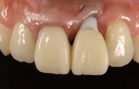
Figure 1 Preoperative intraoral view: 3 mm of vertical abutment exposure was observed.
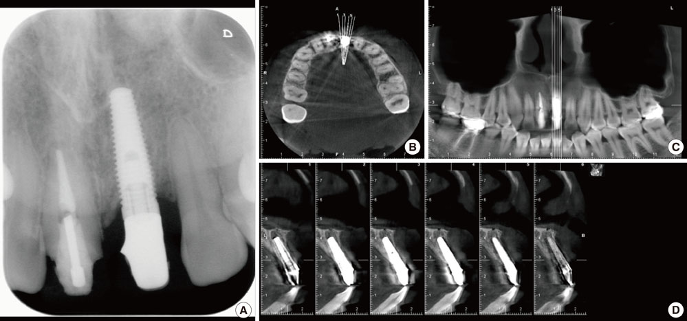
Figure 2 Preoperative radiographical examination of the left maxillary central incisor region: intraoral periapical radiograph (A) and come beam computed tomography (B, axial view; C, coronal view; D, sagittal view).
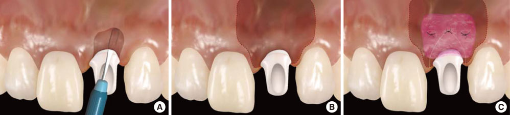
Figure 3 Surgical steps of the subepithelial connective tissue graft (SCTG) with split pouch technique: (A) A circumferential partial-thickness incision was performed using a round tip blade. (B) A partial-thickness pouch was constructed around the dehiscence. (C) The SCTG was positioned and sutured in the pouch with normal tension.
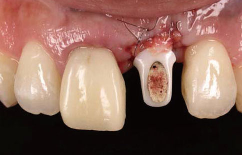
Figure 4 Intraoral appearance immediately after the first subepithelial connective tissue graft.
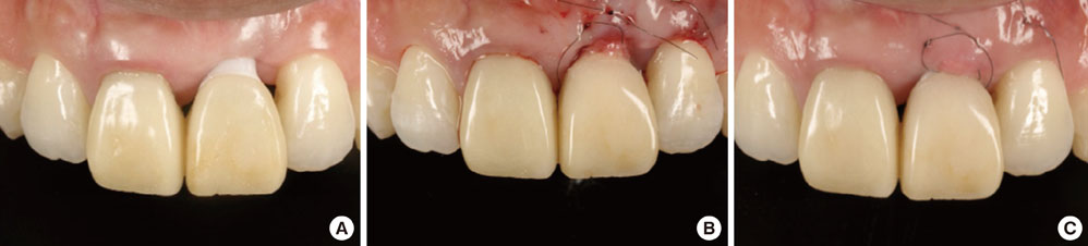
Figure 5 (A) Intraoral appearance at 12 months after the first subepithelial connective tissue graft (SCTG): The mucosal dehiscence was partially covered by epithelium. (B) Intraoperative view in the second stage surgery: The SCTG was positioned and sutured in the pouch with normal tension. (C) Intraoral appearance at 2 weeks after the second SCTG.
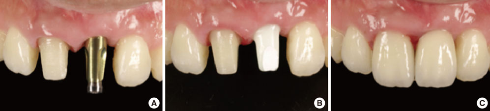
Figure 6 Prosthetic procedures: (A) intraoral view after connection of impression coping, (B) intraoral view after connection of custom zirconia abutment, and (C) intraoral view immediately after placement of all-ceramic crowns.
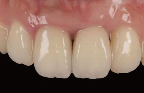
Figure 7 Nine months after the second SCTG. Sufficient soft tissue regeneration was achieved and the marginal soft tissue level was stable and esthetically satisfactory.
Reference
-
1. Berglundh T, Lindhe J. Dimension of the periimplant mucosa. Biological width revisited. J Clin Periodontol. 1996. 23:971–973.2. Cardaropoli G, Lekholm U, Wennstrom JL. Tissue alterations at implant-supported single-tooth replacements: a 1-year prospective clinical study. Clin Oral Implants Res. 2006. 17:165–171.
Article3. Muller HP, Heinecke A, Schaller N, Eger T. Masticatory mucosa in subjects with different periodontal phenotypes. J Clin Periodontol. 2000. 27:621–626.
Article4. Belser UC, Buser D, Hess D, Schmid B, Bernard JP, Lang NP. Aesthetic implant restorations in partially edentulous patients: a critical appraisal. Periodontol 2000. 1998. 17:132–150.
Article5. Burkhardt R, Joss A, Lang NP. Soft tissue dehiscence coverage around endosseous implants: a prospective cohort study. Clin Oral Implants Res. 2008. 19:451–457.
Article6. Roccuzzo M, Bunino M, Needleman I, Sanz M. Periodontal plastic surgery for treatment of localized gingival recessions: a systematic review. J Clin Periodontol. 2002. 29:Suppl 3. 178–194.
Article7. Nozawa T, Enomoto H, Tsurumaki S, Ito K. Biologic height-width ratio of the buccal supra-implant mucosa. Eur J Esthet Dent. 2006. 1:208–214.8. Nabers JM. Free gingival grafts. Periodontics. 1966. 4:243–245.9. Soileau KM, Brannon RB. A histologic evaluation of various stages of palatal healing following subepithelial connective tissue grafting procedures: a comparison of eight cases. J Periodontol. 2006. 77:1267–1273.
Article10. Oates TW, Robinson M, Gunsolley JC. Surgical therapies for the treatment of gingival recession. A systematic review. Ann Periodontol. 2003. 8:303–320.
Article11. Zucchelli G, Mele M, Mazzotti C, Marzadori M, Montebugnoli L, De Sanctis M. Coronally advanced flap with and without vertical releasing incisions for the treatment of multiple gingival recessions: a comparative controlled randomized clinical trial. J Periodontol. 2009. 80:1083–1094.
Article12. Cortellini P, Tonetti M, Baldi C, Francetti L, Rasperini G, Rotundo R, et al. Does placement of a connective tissue graft improve the outcomes of coronally advanced flap for coverage of single gingival recessions in upper anterior teeth? A multi-centre, randomized, double-blind, clinical trial. J Clin Periodontol. 2009. 36:68–79.
Article13. Clauser C, Nieri M, Franceschi D, Pagliaro U, Pini-Prato G. Evidence-based mucogingival therapy. Part 2: Ordinary and individual patient data meta-analyses of surgical treatment of recession using complete root coverage as the outcome variable. J Periodontol. 2003. 74:741–756.
Article14. Miller PD Jr. A classification of marginal tissue recession. Int J Periodontics Restorative Dent. 1985. 5:8–13.
- Full Text Links
-
- Actions
-
Cited
- CITED
-
- Close
- Share
-
- Similar articles
-
- Surgical Approach for Facial Peri-implant Soft Tissue Deficiency in the Esthetic Area
- Endoscope-assisted Removal of Dental Implants Displaced Into the Maxillary Sinus: A Report of 27 Patients
- Tissue integration of zirconia and titanium implants with and without buccal dehiscence defects
- Risk Factors for Wound Dehiscence after Guided Bone Regeneration in Dental Implant Surgery
- Complications after dental implant surgery with iliac bone graft; Case report
