A comparison of different gingival depigmentation techniques: ablation by erbium:yttrium-aluminum-garnet laser and abrasion by rotary instruments
- Affiliations
-
- 1Department of Periodontology, Kyung Hee University School of Dentistry, Seoul, Korea. yherr@khu.ac.kr
- 2Institute of Oral Biology, Kyung Hee University School of Dentistry, Seoul, Korea.
- KMID: 2094720
- DOI: http://doi.org/10.5051/jpis.2011.41.4.201
Abstract
- PURPOSE
The aim of this study is to compare two different gingival depigmentation techniques using an erbium:yttrium-aluminum-garnet (Er:YAG) laser and rotary instruments.
METHODS
Two patients with melanin pigmentation of gingiva were treated with different gingival depigmentation techniques. Ablation of the gingiva by Er:YAG laser was performed on the right side, and abrasion with a rotary round bur on the opposite side.
RESULTS
The patients were satisfied with the esthetically significant improvement with each method. However, some pigment still remained on the marginal gingival and papilla. The visual analog scale did not yield much difference between the two methods, with slightly more pain on the Er:YAG laser treated site.
CONCLUSIONS
The results of these cases suggest that ablation of the gingiva by an Er:YAG laser and abrasion with a rotary round bur is good enough to achieve esthetic satisfaction and fair wound healing without infection or severe pain. Prudent care about the gingival condition, such as the gingival thickness and degree of pigmentation along with appropriate assessment is needed in ablation by the Er:YAG laser procedure.
Keyword
Figure
-
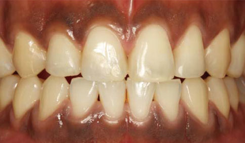
Figure 1 Case 1: pre-operative view. Diffused melanin hyperpigmentation was observed.
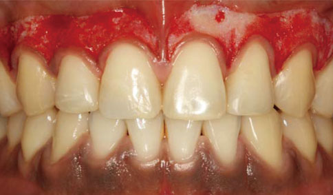
Figure 2 Post-operative view.
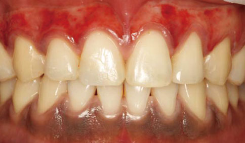
Figure 3 Postoperative view (1 day later). The connective tissue was still exposed.
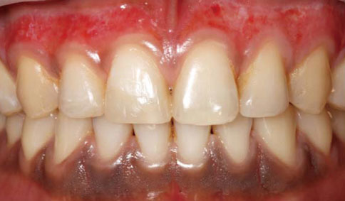
Figure 4 Postoperative view (1 week later).

Figure 5 Postoperative view (2 weeks later). Re-epithelization was almost complete but the epithelium had not yet recovered its thickness.
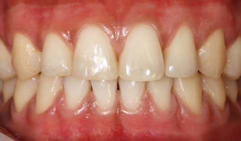
Figure 6 Postoperative view (upper jaw, 4 weeks later; lower jaw, 2 weeks later).
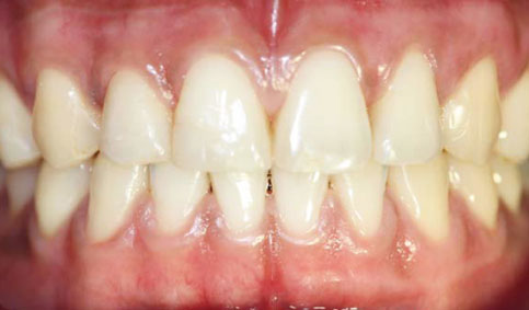
Figure 7 Postoperative view (upper jaw, 5 weeks later; lower jaw, 3 weeks later). The gingiva showed a normal appearance with a pink color and keratinization.

Figure 8 Case 2: preoperative view. The patient showed focal pigmentation.
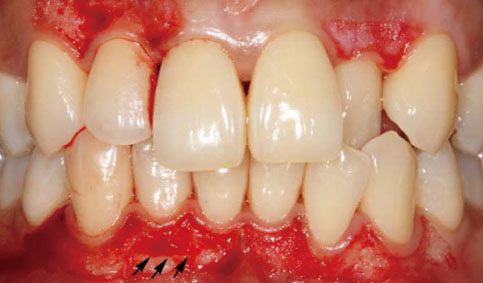
Figure 9 Postoperative view. Exposure of underlying bone can be observed around the incisal area on the right mandible. Arrows show the exposed area.

Figure 10 Postoperative view (1 week later). Unfavorable healing is observed with the appearance of necrosis on the right side of the mandibular area.

Figure 11 Postoperative view (2 weeks later).

Figure 12 Post-operative view (4 weeks later). The wound healing was almost completed, but slight redness remained.
Reference
-
1. Kauzman A, Pavone M, Blanas N, Bradley G. Pigmented lesions of the oral cavity: review, differential diagnosis, and case presentations. J Can Dent Assoc. 2004. 70:682–683.2. Mirowski GW, Waibel JS. Pigmented lesions of the oral cavity. Dermatol Ther. 2002. 15:218–228.
Article3. Meleti M, Vescovi P, Mooi WJ, van der Waal I. Pigmented lesions of the oral mucosa and perioral tissues: a flow-chart for the diagnosis and some recommendations for the management. Oral Surg Oral Med Oral Pathol Oral Radiol Endod. 2008. 105:606–616.
Article4. Hedin CA, Pindborg JJ, Axéll T. Disappearance of smoker's melanosis after reducing smoking. J Oral Pathol Med. 1993. 22:228–230.
Article5. Hedin CA, Axéll T. Oral melanin pigmentation in 467 Thai and Malaysian people with special emphasis on smoker's melanosis. J Oral Pathol Med. 1991. 20:8–12.
Article6. Araki S, Murata K, Ushio K, Sakai R. Dose-response relationship between tobacco consumption and melanin pigmentation in the attached gingiva. Arch Environ Health. 1983. 38:375–378.
Article7. Eisen D. Disorders of pigmentation in the oral cavity. Clin Dermatol. 2000. 18:579–587.8. Perlmutter S, Tal H. Repigmentation of the gingiva following surgical injury. J Periodontol. 1986. 57:48–50.
Article9. Farnoosh AA. Treatment of gingival pigmentation and discoloration for esthetic purposes. Int J Periodontics Restorative Dent. 1990. 10:312–319.10. Tamizi M, Taheri M. Treatment of severe physiologic gingival pigmentation with free gingival autograft. Quintessence Int. 1996. 27:555–558.11. Tal H, Landsberg J, Kozlovsky A. Cryosurgical depigmentation of the gingiva. A case report. J Clin Periodontol. 1987. 14:614–617.
Article12. Yeh CJ. Cryosurgical treatment of melanin-pigmented gingiva. Oral Surg Oral Med Oral Pathol Oral Radiol Endod. 1998. 86:660–663.
Article13. Arikan F, Gürkan A. Cryosurgical treatment of gingival melanin pigmentation with tetrafluoroethane. Oral Surg Oral Med Oral Pathol Oral Radiol Endod. 2007. 103:452–457.
Article14. Pontes AE, Pontes CC, Souza SL, Novaes AB Jr, Grisi MF, Taba M Jr. Evaluation of the efficacy of the acellular dermal matrix allograft with partial thickness flap in the elimination of gingival melanin pigmentation. A comparative clinical study with 12 months of follow-up. J Esthet Restor Dent. 2006. 18:135–143.
Article15. Nakamura Y, Hossain M, Hirayama K, Matsumoto K. A clinical study on the removal of gingival melanin pigmentation with the CO(2) laser. Lasers Surg Med. 1999. 25:140–147.
Article16. Atsawasuwan P, Greethong K, Nimmanon V. Treatment of gingival hyperpigmentation for esthetic purposes by Nd:YAG laser: report of 4 cases. J Periodontol. 2000. 71:315–321.
Article17. Tal H, Oegiesser D, Tal M. Gingival depigmentation by erbium:YAG laser: clinical observations and patient responses. J Periodontol. 2003. 74:1660–1667.
Article18. Rosa DS, Aranha AC, Eduardo Cde P, Aoki A. Esthetic treatment of gingival melanin hyperpigmentation with Er:YAG laser: short-term clinical observations and patient follow-up. J Periodontol. 2007. 78:2018–2025.
Article19. Azzeh MM. Treatment of gingival hyperpigmentation by erbium-doped:yttrium, aluminum, and garnet laser for esthetic purposes. J Periodontol. 2007. 78:177–184.
Article20. Aoki A, Sasaki KM, Watanabe H, Ishikawa I. Lasers in nonsurgical periodontal therapy. Periodontol 2000. 2004. 36:59–97.
Article21. Ishikawa I, Sasaki KM, Aoki A, Watanabe H. Effects of Er:YAG laser on periodontal therapy. J Int Acad Periodontol. 2003. 5:23–28.22. Watanabe H, Ishikawa I, Suzuki M, Hasegawa K. Clinical assessments of the erbium:YAG laser for soft tissue surgery and scaling. J Clin Laser Med Surg. 1996. 14:67–75.
Article23. Berger JW, D'Amico DJ. Modeling of erbium: YAG laser-mediated explosive photovaporization: implications for vitreoretinal surgery. Ophthalmic Surg Lasers. 1997. 28:133–139.
Article24. Ando Y, Aoki A, Watanabe H, Ishikawa I. Bactericidal effect of erbium YAG laser on periodontopathic bacteria. Lasers Surg Med. 1996. 19:190–200.
Article25. Ozcelik O, Cenk Haytac M, Kunin A, Seydaoglu G. Improved wound healing by low-level laser irradiation after gingivectomy operations: a controlled clinical pilot study. J Clin Periodontol. 2008. 35:250–254.
Article26. Qadri T, Miranda L, Tunér J, Gustafsson A. The short-term effects of low-level lasers as adjunct therapy in the treatment of periodontal inflammation. J Clin Periodontol. 2005. 32:714–719.
Article27. Esen E, Haytac MC, Oz IA, Erdoğan O, Karsli ED. Gingival melanin pigmentation and its treatment with the CO2 laser. Oral Surg Oral Med Oral Pathol Oral Radiol Endod. 2004. 98:522–527.
Article
- Full Text Links
-
- Actions
-
Cited
- CITED
-
- Close
- Share
-
- Similar articles
-
- Erbium-doped Yttrium Aluminum Garnet Laser to Treat Familial Acanthosis Nigricans
- A Pilot Study of Skin Resurfacing Using the 2,790-nm Erbium:YSGG Laser System
- Erbium: YAG Laser Sclerostomy in Rabbits
- Acquired Dermal Melanocytosis of the Nose Successfully Treated with Neodymium-doped Yttrium Aluminum Garnet 1064 nm Laser
- A Prospective Split-Face Comparative Study of Periorbital Wrinkle Treatments: Fractional Erbium-Doped Yttrium Aluminum Garnet Laser, Intense Pulsed Light, and Topical 0.1% Tretinoin Cream
