Korean J Hematol.
2006 Jun;41(2):124-128. 10.5045/kjh.2006.41.2.124.
A Case of Primary Hepatic B-cell Lymphoma of Mucosa-associated Lymphoid Tissue (MALT)-Type
- Affiliations
-
- 1Department of Internal Medicine, Fatima Hospital, Daegu, Korea.
- 2Department of Internal Medicine, Fatima Hospital, Changwon, Korea. jshbjh@yahoo.co.kr
- 3Department of Pathology, Fatima Hospital, Changwon, Korea.
- 4Department of General Surgery, Fatima Hospital, Changwon, Korea.
- KMID: 2083488
- DOI: http://doi.org/10.5045/kjh.2006.41.2.124
Abstract
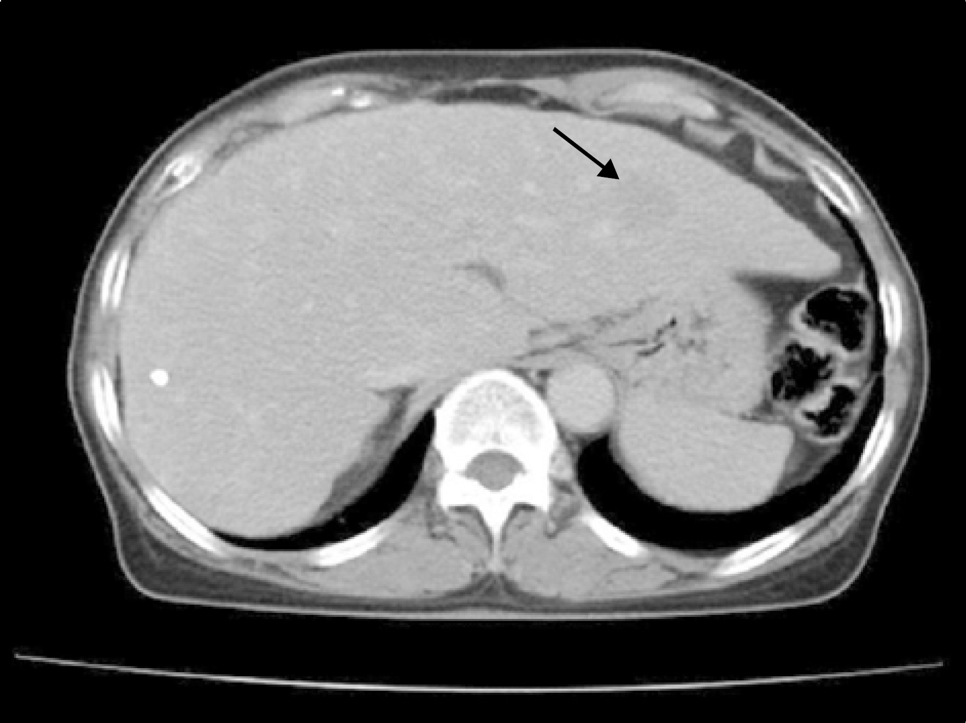
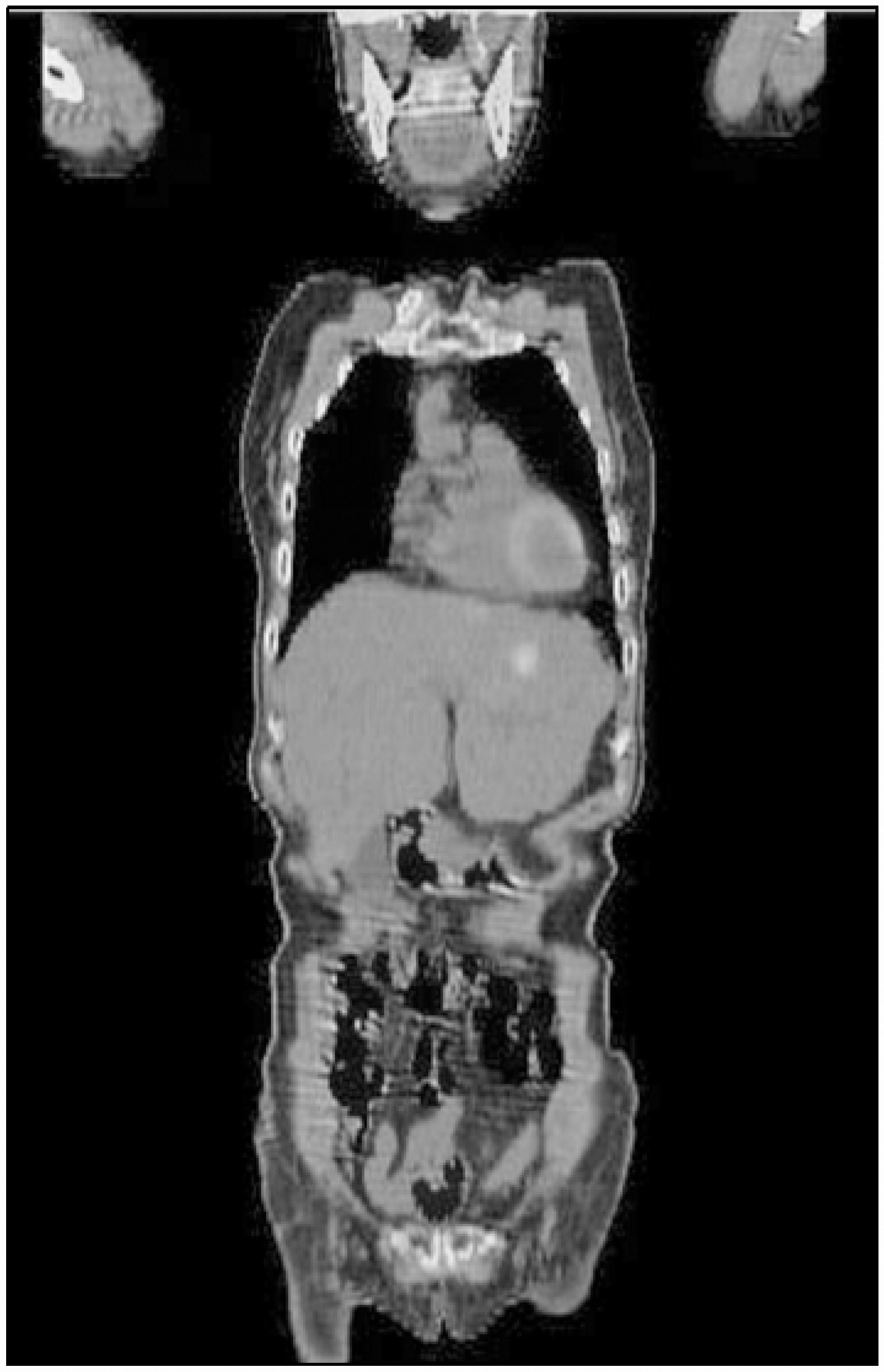
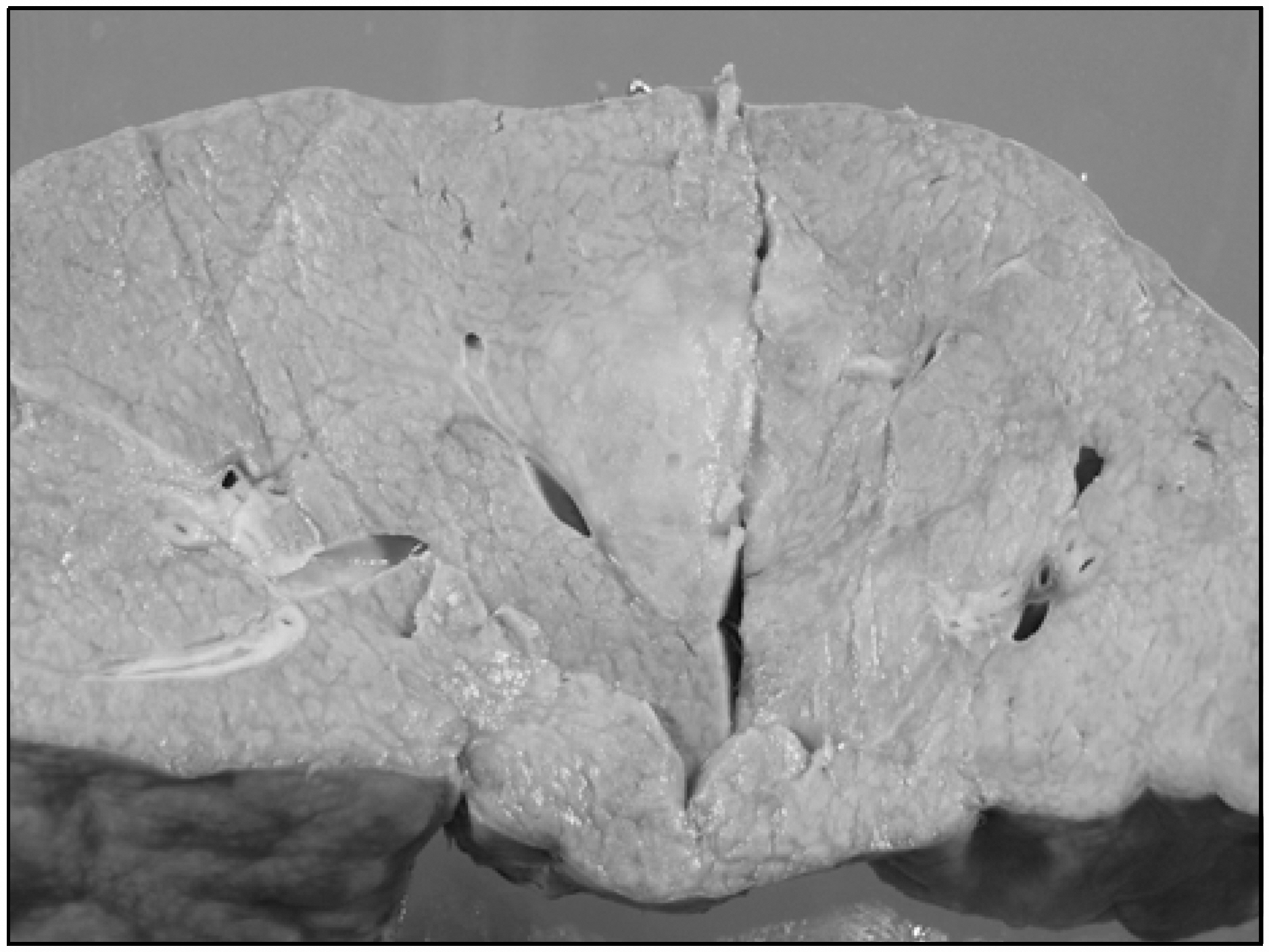
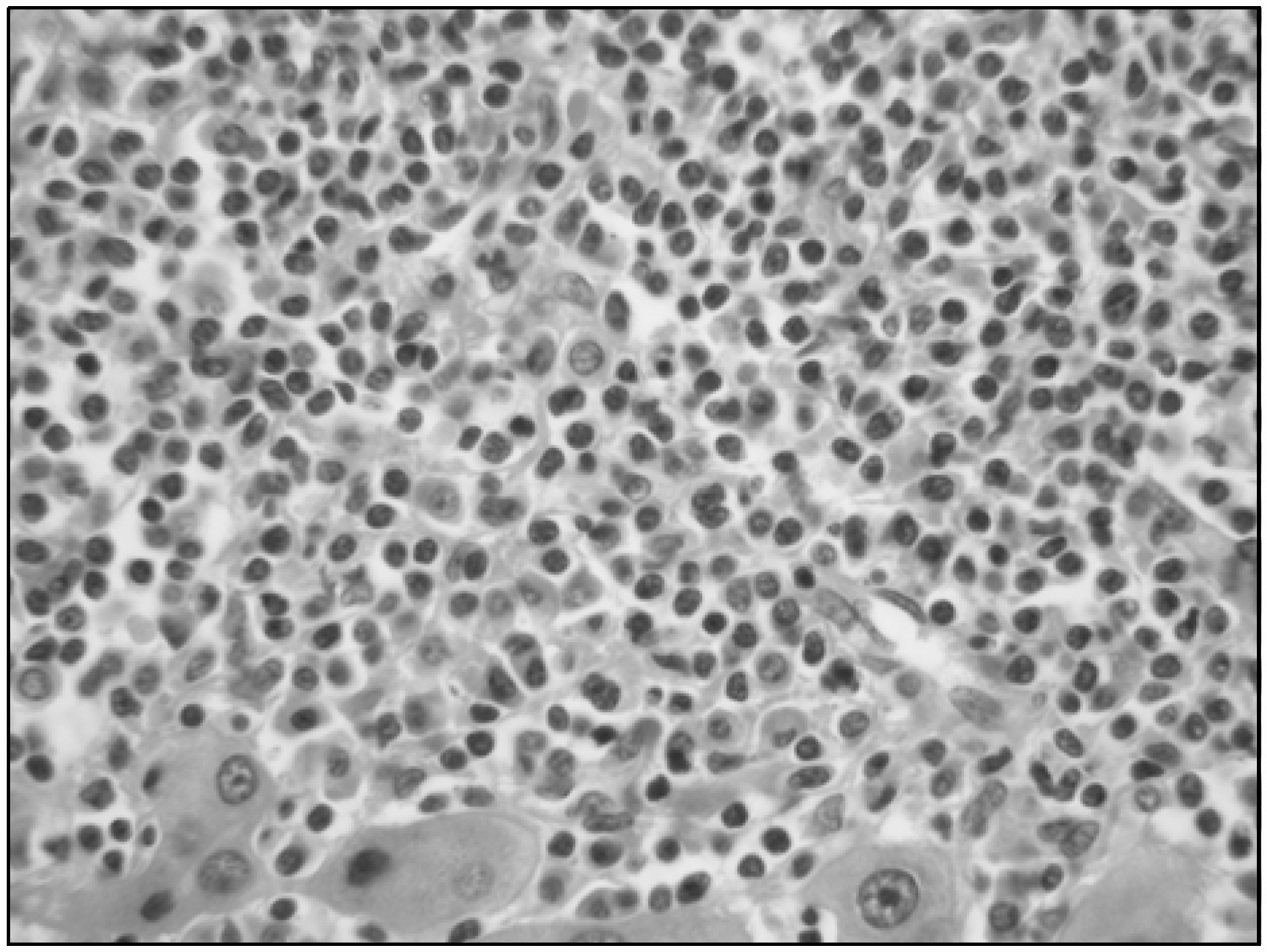
- Mucosa-associated lymphoid tissue (MALT) lymphoma is a low grade B cell lymphoma that, occurs in numerous sites including the stomach, ocular adnexa, thyroid, lung and breast; however, primary hepatic lymphoma is extremely rare. Only about 20 cases have been reported world wide. We recently experienced a case of primary hepatic B-cell lymphoma of the MALT type in a 63-year old female patient. She presented with abdominal pain. The CT, ultrasonogram and PET-CT showed a hepatic nodular mass. A biopsy specimen of the liver revealed MALT lymphoma. There was no evidence of the lymphoma in the extrahepatic lesion. She received segmentectomy of liver and was then treated with CVP (cyclophosphamide, vincristine and prednisolone) chemotherapy. She has been followed up for 6 months since the therapy, and she remains asymptomatic.
Keyword
MeSH Terms
Figure
Reference
-
1). Isaacson P., Wright DH. Malignant lymphoma of mucosa-associated lymphoid tissue. A distinctive type of B-cell lymphoma. Cancer. 1983. 52:1410–6.
Article2). Harris NL., Jaffe ES., Stein H, et al. A revised european-american classification of lymphoid neoplasms: a proposal from the International Lymphoma Study Group. Blood. 1994. 84:1361–92.3). Harris NL., Jaffe ES., Diebold J, et al. World Health Organization Classification of Neoplastic Diseases of the hematopoietic and lymphoid tissues: report of the Clinical Advisory Committee Meeting-Airlie House, Virginia. J Clin Oncol. 1999. 17:3835–49.4). Zinzani PL., Magagnoli M., Galieni P, et al. Nongastr-ointestinal low-grade mucosa-associated lymphoid tissue lymphoma: analysis of 75 patients. J Clin Oncol. 1999. 17:1254.
Article5). Lei KI. Primary non-hodgkin's lymphoma of the liver. Leuk Lymphoma. 1998. 29:293–9.6). Yang US., Cho M., Song CS, et al. A case of primary low-grade hepatic b-cell lymphoma of mucosa-associated lymphoid tissue (MALT)-type. Korean J Gas-troenterol. 1998. 31:547–52.7). Noronha V., Shafi NQ., Obando JA., Kummar S. Primary non-Hodgkin's lymphoma of the liver. Crit Rev Oncol Hematol. 2005. 53:199–207.
Article8). Avlonitis VS., Linos D. Primary hepatic lymphoma: a review. Eur J Surg. 1999. 165:725–9.9). Levy AD. Malignant liver tumors. Clin Liver Dis. 2002. 6:147–64.
Article10). Rizzi EB., Schinina V., Cristofaro M., David V., Bibboli-no C. Non-Hodgkin's lymphoma of the liver in patients with AIDS: sonographic, CT and MRI findings. J Clin Ultrasound. 2001. 29:125–9.11). Elstrom R., Guan L., Baker G, et al. Utility of FDG-PET Scanning in lymphoma by WHO classification. Blood. 2003. 15:3875–6.
Article12). Hyjek E., Isaacson PG. Primary B-cell lymphoma of the thyroid and its relationship to Hashimoto's thyroiditis. Hum Pathol. 1998. 19:1315–26.13). Isaacson PG., Banks PM., Best PV., McLure SP., Muller-Hermelink HK., Wyatt JI. Primary low-grade hepatic B-cell lymphoma of mucosa-associated lymphoid tissue (MALT)-type. Am J Surg Pathol. 1995. 19:571–5.
Article14). Kirk CM., Lewin D., Lazarchick J. Primary hepatic b-cell lymphoma of mucosa-associated lymphoid tissue. Arch Pathol Lab Med. 1999. 123:716–9.
Article15). Tsang RW., Gospodarowicz MK., Pintilie M, et al. Localized mucosa-associated lymphoid tissue lymphoma treated with radiation therapy has excellent clinical outcome. J Clin Oncol. 2003. 21:4157–64.
Article16). Klasa RJ., Meyer RM., Shustik C, et al. Randomized phase III study of fludarabine phosphate versus cyclophosphamide, vincristine, and prednisone in patients with recurrent low grade non Hodgkin's lymphoma previously treated with an alkylating agent or alkylator-containing regimen. J Clin Oncol. 2002. 20:4649–54.
- Full Text Links
- Actions
-
Cited
- CITED
-
- Close
- Share
- Similar articles
-
- Longlasting Remission of Primary Hepatic Mucosa-associated Lymphoid Tissue (MALT) Lymphoma Achieved by Radiotherapy Alone
- A case report of the Pulmonary Malignant Lymphomaof the mucosa-associated lymphoid tissue(MALT)
- A Case of Primary Pulmonary Extranodal Marginal Zone B-Cell Lymphoma of the MALT Type
- Role of Chemotherapy in Gastric Marginal Zone B-Cell Lymphoma of Mucosa-Associated Lymphoid Tissue (MALT) Type
- Primary Mucosa-Associated Lymphoid Tissue Lymphoma of the Breast with Synchronous Contralateral Invasive Breast Cancer: A Case Report

