Uncommon Manifestations of Scrub Typhus Encephalitis in Two Cases: Clinical and Magnetic Resonance Imaging Findings
- Affiliations
-
- 1Department of Radiology, Inje University Busan Paik Hospital, Busan, Korea. hwjeong2000@lycos.co.kr
- KMID: 2079570
- DOI: http://doi.org/10.3348/jksr.2015.73.5.337
Abstract
- Scrub typhus is a well-known acute febrile illness caused by Orientia tsutsugamushi. This disease has multiorgan involvement, which includes the lungs, heart, liver, spleen, and the central or peripheral nervous system. Scrub typhus involving the central nervous system (CNS) is not rare. However, meningitis and meningoencephalitis can cause changes in mentation and death and are therefore associated with a poor prognosis. We report two consecutive cases of scrub typhus with CNS involvement. One patient presented with extensive white matter involvement, similar to that observed in acute disseminated encephalomyelitis, whereas the other patient presented with subependymal enhancement along the lateral ventricles. To the best of our knowledge, scrub typhus encephalitis, with extensive white matter involvement and subependymal enhancement, are very rarely described findings in the previous literature. Our patients did not show complete recovery, but the symptoms resolved with treatment. Recognizing these uncommon radiologic findings of scrub typhus may be helpful in the early diagnosis of scrub typhus with CNS involvement, which may alter the prognoses of patients.
MeSH Terms
Figure
-
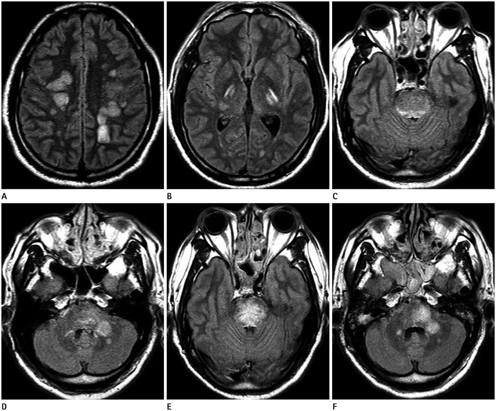
Fig. 1 Magnetic resonance imaging in a 57-year-old man with general weakness, gait disturbance, and altered mental status. Axial fluid-attenuated inversion recovery images show multifocal, hyperintense lesions involving the cerebral white matter on both the right and left sides (A), thalamus (B), brainstem (C), left middle cerebellar peduncle, and both cerebellar hemispheres (D). A follow-up magnetic resonance image obtained 2 weeks later shows an increased extent of hyperintense lesions involving the brainstem, and no significant interval change in the lesions involving the left middle cerebellar peduncle (E) and both cerebellar hemispheres (F).

Fig. 2 Magnetic resonance imaging in a 71-year-old woman with general weakness. Contrast-enhanced T1-weighted images show focal enhancement involving the right gyrus rectus (arrow) (A) and subtle subependymal enhancement (B, C) along both lateral ventricles (arrows). After 10 days of persistent medical therapy, a coronal contrast-enhanced T1-weighted image shows that the subependymal enhancement along the both the lateral ventricles (arrows) has disappeared (D), and axial fluid-attenuated inversion recovery images show new multifocal hyperintense lesions in both the basal ganglia and right frontal lobe (arrows) (E).
Reference
-
1. Kim HC, Yoon KW, Yoo DS, Cho CS. Hemorrhagic Transformation of Scrub Typhus Encephalitis: A Rare Entity. Clin Neuroradiol. 2014; 11. 06. [Epub].2. Chua CJ, Tan KS, Ramli N, Devi S, Tan CT. Scrub typhus with central nervous system involvement: a case report with CT and MR imaging features. Neurol J Southeast Asia. 1999; 4:53–57.3. Kim DE, Lee SH, Park KI, Chang KH, Roh JK. Scrub typhus encephalomyelitis with prominent focal neurologic signs. Arch Neurol. 2000; 57:1770–1772.4. Sood S, Sharma S, Khanna S. Role of advanced MRI brain sequences in diagnosing neurological complications of scrub typhus. J Clin Imaging Sci. 2015; 5:11.5. Choi YH, Kim SJ, Lee JY, Pai HJ, Lee KY, Lee YS. Scrub typhus: radiological and clinical findings. Clin Radiol. 2000; 55:140–144.6. Viswanathan S, Muthu V, Iqbal N, Remalayam B, George T. Scrub typhus meningitis in South India--a retrospective study. PLoS One. 2013; 8:e66595.7. Yum KS, Na SJ, Lee KO, Ko JH. Scrub typhus meningo-encephalitis with focal neurologic signs and associated brain MRI abnormal findings: literature review. Clin Neurol Neurosurg. 2011; 113:250–253.8. Raoult D, Drancourt M. Antimicrobial therapy of rickettsial diseases. Antimicrob Agents Chemother. 1991; 35:2457–2462.9. Kar A, Dhanaraj M, Dedeepiya D, Harikrishna K. Acute encephalitis syndrome following scrub typhus infection. Indian J Crit Care Med. 2014; 18:453–455.10. Jeong YJ, Kim S, Wook YD, Lee JW, Kim KI, Lee SH. Scrub typhus: clinical, pathologic, and imaging findings. Radiographics. 2007; 27:161–172.
- Full Text Links
-
- Actions
-
Cited
- CITED
-
- Close
- Share
-
- Similar articles
-
- A case of scrub typhus with reversible hearing loss
- Opsoclonus-Myoclonus Syndrome Associated with Scrub Typhus
- Facial diplegia as a delayed complication of scrub typhus
- Antibiotic Combination Therapy for Severe Scrub Typhus: Is It Necessary?
- Myeloneuropathy with Bilateral Foot Pain after Scrub Typhus Infection: An Antibody-Proven Case Report
