Korean J Pain.
2014 Apr;27(2):162-167. 10.3344/kjp.2014.27.2.162.
The Radiation Exposure of Radiographer Related to the Location in C-arm Fluoroscopy-guided Pain Interventions
- Affiliations
-
- 1Department of Anesthesiology and Pain Medicine, Konkuk University Medical Center, Seoul, Korea. painfree@kuh.ac.kr
- KMID: 2074063
- DOI: http://doi.org/10.3344/kjp.2014.27.2.162
Abstract
- BACKGROUND
Although a physician may be the nearest to the radiation source during C-arm fluoroscope-guided interventions, the radiographer is also near the fluoroscope. We prospectively investigated the radiation exposure of radiographers relative to their location.
METHODS
The effective dose (ED) was measured with a digital dosimeter on the radiographers' left chest and the side of the table. We observed the location of the radiographers in each procedure related to the mobile support structure of the fluoroscope (Groups A, M and P). Data about age, height, weight, sex, exposure time, radiation absorbed dose (RAD), and the ED at the radiographer's chest and the side of the table was collected.
RESULTS
There were 51 cases for Group A, 116 cases for Group M and 144 cases for Group P. No significant differences were noted in the demographic data such as age, height, weight, and male to female ratio, and exposure time, RAD and ED at the side of the table. Group P had the lowest ED (0.5 +/- 0.8 microSv) of all the groups (Group A, 1.6 +/- 2.3 microSv; Group M, 1.3 +/- 1.9 microSv; P < 0.001). The ED ratio (ED on the radiographer's chest/ED at the side of the table) of Group A was the highest, and the ED radio of Group P was the lowest of all the groups (Group A, 12.2 +/- 21.5%; Group M, 5.7 +/- 6.5%; Group P, 2.5 +/- 6.7%; P < 0.001).
CONCLUSIONS
Radiographers can easily reduce their radiation exposure by changing their position. Two steps behind the mobile support structure can effectively decrease the exposure of radiographers by about 80%.
MeSH Terms
Figure
-
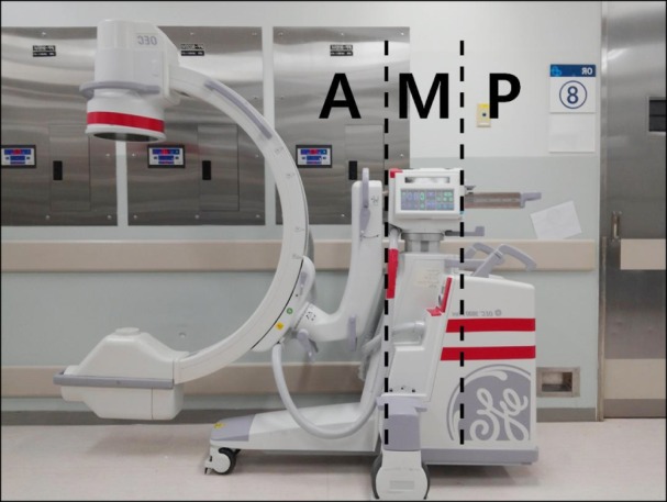
Fig. 1 The Group A, M and P according to the location of dosimeter at radiographer's left chest.
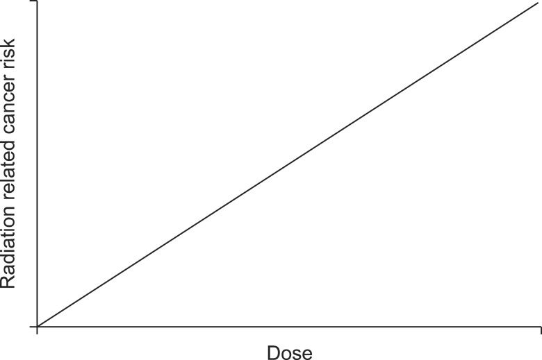
Fig. 2 Graph of linear no threshold model.
Cited by 2 articles
-
Three principles for radiation safety: time, distance, and shielding
Jae Hun Kim
Korean J Pain. 2018;31(3):145-146. doi: 10.3344/kjp.2018.31.3.145.Radiation safety for pain physicians: principles and recommendations
Sewon Park, Minjung Kim, Jae Hun Kim
Korean J Pain. 2022;35(2):129-139. doi: 10.3344/kjp.2022.35.2.129.
Reference
-
1. Baek SW, Ryu JS, Jung CH, Lee JH, Kwon WK, Woo NS, et al. A randomized controlled trial about the levels of radiation exposure depends on the use of collimation C-arm fluoroscopic-guided medial branch block. Korean J Pain. 2013; 26:148–153. PMID: 23614076.
Article2. Ryu JS, Baek SW, Jung CH, Cho SJ, Jung EG, Kim HK, et al. The survey about the degree of damage of radiation-protective shields in operation room. Korean J Pain. 2013; 26:142–147. PMID: 23614075.
Article3. Fish DE, Kim A, Ornelas C, Song S, Pangarkar S. The risk of radiation exposure to the eyes of the interventional pain physician. Radiol Res Pract. 2011; 2011:609537. PMID: 22091381.
Article4. Botwin KP, Freeman ED, Gruber RD, Torres-Rames FM, Bouchtas CG, Sanelli JT, et al. Radiation exposure to the physician performing fluoroscopically guided caudal epidural steroid injections. Pain Physician. 2001; 4:343–348. PMID: 16902680.5. Manchikanti L, Cash KA, Moss TL, Pampati V. Radiation exposure to the physician in interventional pain management. Pain Physician. 2002; 5:385–393. PMID: 16886017.
Article6. Jung CH, Ryu JS, Baek SW, Oh JH, Woo NS, Kim HK, et al. Radiation exposure of the hand and chest during C-arm fluoroscopy-guided procedures. Korean J Pain. 2013; 26:51–56. PMID: 23342208.
Article7. Cho JH, Kim JY, Kang JE, Park PE, Kim JH, Lim JA, et al. A study to compare the radiation absorbed dose of the C-arm fluoroscopic modes. Korean J Pain. 2011; 24:199–204. PMID: 22220241.
Article8. National Council on Radiation Protection and Measurements (US). Occupational dose limits. In: NCRP Report No. 116. Limitation of exposure to ionizing radiation. Bethesda (MD): National Council on Radiation Protection and Measurements;1993. p. 33–35.9. Park PE, Park JM, Kang JE, Cho JH, Cho SJ, Kim JH, et al. Radiation safety and education in the applicants of the final test for the expert of pain medicine. Korean J Pain. 2012; 25:16–21. PMID: 22259711.
Article10. Fink GE. Radiation safety in fluoroscopy for neuraxial injections. AANA J. 2009; 77:265–269. PMID: 19731844.11. Little MP. Do non-targeted effects increase or decrease low dose risk in relation to the linear-non-threshold (LNT) model? Mutat Res. 2010; 687:17–27. PMID: 20105434.
Article12. Shah DJ, Sachs RK, Wilson DJ. Radiation-induced cancer: a modern view. Br J Radiol. 2012; 85:e1166–e1173. PMID: 23175483.
Article13. Hendee WR, O'Connor MK. Radiation risks of medical imaging: separating fact from fantasy. Radiology. 2012; 264:312–321. PMID: 22821690.
Article14. Crawley MT, Shine B, Booth A. Radiation dose and diagnosticity of barium enema examinations by radiographers and radiologists: a comparative study. Br J Radiol. 1998; 71:399–405. PMID: 9659133.
Article15. Tilson ER. Educational and experiential effects on radiographers' radiation safety behavior. Radiol Technol. 1982; 53:321–325. PMID: 7063661.16. Johnston J, Killion JB, Vealé B, Comello R. U.S. Technologists' radiation exposure perceptions and practices. Radiol Technol. 2011; 82:311–320. PMID: 21406708.17. Vano E, Gonzalez L, Fernandez JM, Prieto C, Guibelalde E. Influence of patient thickness and operation modes on occupational and patient radiation doses in interventional cardiology. Radiat Prot Dosimetry. 2006; 118:325–330. PMID: 16439516.
Article18. Ghobadifar MA, Zarei S. Effect of collimation on radiation exposure and image quality. Korean J Pain. 2013; 26:307–308. PMID: 23862008.
Article19. Miller DL, Vañó E, Bartal G, Balter S, Dixon R, Padovani R, et al. Occupational radiation protection in interventional radiology: a joint guideline of the Cardiovascular and Interventional Radiology Society of Europe and the Society of Interventional Radiology. Cardiovasc Intervent Radiol. 2010; 33:230–239. PMID: 20020300.
Article
- Full Text Links
-
- Actions
-
Cited
- CITED
-
- Close
- Share
-
- Similar articles
-
- Radiation safety for pain physicians: principles and recommendations
- Radiation exposure and protection for eyes in pain management
- Factors Related to Radiation Exposure during Lumbar Spine Intervention
- Radiation safety: a focus on lead aprons and thyroid shields in interventional pain management
- How Effective Are Radiation Reducing Gloves in C-arm Fluoroscopy-guided Pain Interventions?
