How Effective Are Radiation Reducing Gloves in C-arm Fluoroscopy-guided Pain Interventions?
- Affiliations
-
- 1Department of Anesthesiology and Pain Medicine, Konkuk University Medical Center, Seoul, Korea. painfree@kuh.ac.kr
- KMID: 2074061
- DOI: http://doi.org/10.3344/kjp.2014.27.2.145
Abstract
- BACKGROUND
The physician's hands are close to the X-ray field in C-arm fluoroscopy-guided pain interventions. We prospectively investigated the radiation attenuation of Proguard RR-2 gloves.
METHODS
In 100 cases, the effective doses (EDs) of two dosimeters without a radiation-reducing glove were collected. EDs from the two dosimeters-one dosimeter wrapped with a glove and the other dosimeter without a glove- were also measured at the side of the table (Group 1, 140 cases) and at a location 20 cm away from the side of the table (Group 2, 120 cases). Mean differences such as age, height, weight, radiation absorbed dose (RAD), exposure time, ED, and ratio of EDs were analyzed.
RESULTS
In the EDs of two dosimeters without gloves, there were no significant differences (39.0 +/- 36.3 microSv vs. 38.8 +/- 36.4 microSv) (P = 0.578). The RAD (192.0 +/- 182.0 radcm2) in Group 2 was higher than that (132.3 +/- 103.5 radcm2) in Group 1 (P = 0.002). The ED (33.3 +/- 30.9 microSv) of the dosimeter without a glove in Group 1 was higher than that (12.3 +/- 8.8 microSv) in Group 2 (P < 0.001). The ED (24.4 +/- 22.4 microSv) of the dosimeter wrapped with a glove in Group 1 was higher than that (9.2 +/- 6.8 microSv) in Group 2 (P < 0.001). No significant differences were noted in the ratio of EDs (73.5 +/- 6.7% vs. 74.2 +/- 9.3%, P = 0.469) between Group 1 and Group 2.
CONCLUSIONS
Proguard RR-2 gloves have a radiation attenuation effect of 25.8-26.5%. The radiation attenuation is not significantly different by intensity of scatter radiation or the different RADs of C-arm fluoroscopy.
Keyword
MeSH Terms
Figure
-
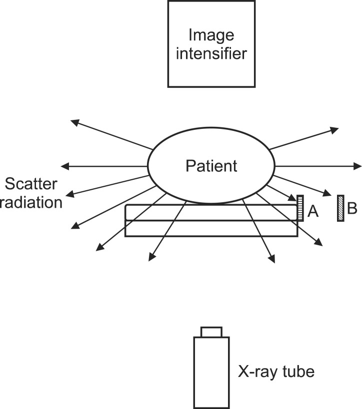
Fig. 1 Study diagram and position of dosimeters in each group (arrow: scatter radiation). A: position of dosimeters at side of table. B: position of dosimeters at 20 cm away from side of table.
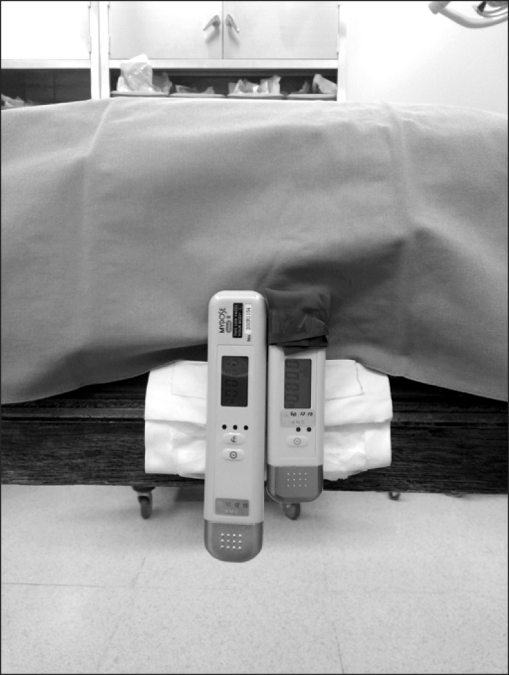
Fig. 2 The position of the two dosimeters at the side of the table. One dosimeter was wrapped with a radiation-reducing glove. Only one layer of the piece totally covered the radiation sensor of the dosimeter. Sensors were facing the patient and table.
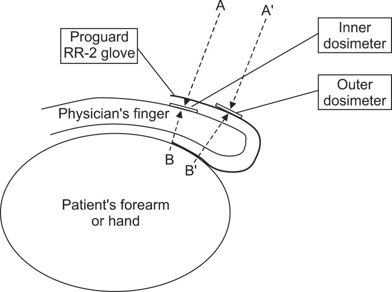
Fig. 3 Schematic diagram of Calder et al.'s [7] study (dotted arrow: scatter radiation). A: attenuated scatter radiation through one layer of radiation-reducing gloves. B: attenuated scatter radiation through one layer of radiation-reducing glove and physician's finger. A': direct scatter radiation. B': attenuated scatter radiation through two layers of radiation-reducing glove and physician's finger.
Cited by 4 articles
-
Radiation Safety for Pain Physicians: Technique or Equipment
Jae Hang Shim
Korean J Pain. 2014;27(2):101-102. doi: 10.3344/kjp.2014.27.2.101.The radiation safety education and the pain physicians' efforts to reduce radiation exposure
Tae Hee Kim, Seung Wan Hong, Nam Sik Woo, Hae Kyoung Kim, Jae Hun Kim
Korean J Pain. 2017;30(2):104-115. doi: 10.3344/kjp.2017.30.2.104.Three principles for radiation safety: time, distance, and shielding
Jae Hun Kim
Korean J Pain. 2018;31(3):145-146. doi: 10.3344/kjp.2018.31.3.145.Radiation safety for pain physicians: principles and recommendations
Sewon Park, Minjung Kim, Jae Hun Kim
Korean J Pain. 2022;35(2):129-139. doi: 10.3344/kjp.2022.35.2.129.
Reference
-
1. Park PE, Park JM, Kang JE, Cho JH, Cho SJ, Kim JH, et al. Radiation safety and education in the applicants of the final test for the expert of pain medicine. Korean J Pain. 2012; 25:16–21. PMID: 22259711.
Article2. Kim C, Vasaiwala S, Haque F, Pratap K, Vidovich MI. Radiation safety among cardiology fellows. Am J Cardiol. 2010; 106:125–128. PMID: 20609659.
Article3. Jung CH, Ryu JS, Baek SW, Oh JH, Woo NS, Kim HK, et al. Radiation exposure of the hand and chest during C-arm fluoroscopy-guided procedures. Korean J Pain. 2013; 26:51–56. PMID: 23342208.
Article4. Fink GE. Radiation safety in fluoroscopy for neuraxial injections. AANA J. 2009; 77:265–269. PMID: 19731844.5. Schueler BA. Operator shielding: how and why. Tech Vasc Interv Radiol. 2010; 13:167–171. PMID: 20723831.
Article6. Ryu JS, Baek SW, Jung CH, Cho SJ, Jung EG, Kim HK, et al. The survey about the degree of damage of radiation-protective shields in operation room. Korean J Pain. 2013; 26:142–147. PMID: 23614075.
Article7. Calder PR, Tennent TD, Allen PW. Assessment of the efficacy of Proguard RR-2 radio-protective gloves during forearm manipulation. Injury. 2003; 34:159–161. PMID: 12565026.
Article8. Mori H, Koshida K, Ishigamori O, Matsubara K. Evaluation of the effectiveness of X-ray protective aprons in experimental and practical fields. Radiol Phys Technol. 2014; 7:158–166. PMID: 24338033.
Article9. Arnstein PM, Richards AM, Putney R. The risk from radiation exposure during operative X-ray screening in hand surgery. J Hand Surg Br. 1994; 19:393–396. PMID: 8077836.
Article10. Koukorava C, Carinou E, Simantirakis G, Vrachliotis TG, Archontakis E, Tierris C, et al. Doses to operators during interventional radiology procedures: focus on eye lens and extremity dosimetry. Radiat Prot Dosimetry. 2011; 144:482–486. PMID: 21044993.
Article11. Baek SW, Ryu JS, Jung CH, Lee JH, Kwon WK, Woo NS, et al. A randomized controlled trial about the levels of radiation exposure depends on the use of collimation C-arm fluoroscopic-guided medial branch block. Korean J Pain. 2013; 26:148–153. PMID: 23614076.
Article12. Hernández García JM, Vidal Marcos A, Gasco García C. A survey on the use of fluoroscopy in the treatment of pain: do we perform it correctly? Rev Esp Anestesiol Reanim. 2012; 59:430–435. PMID: 22824536.13. Rehani MM, Ciraj-Bjelac O, Vañó E, Miller DL, Walsh S, Giordano BD, et al. ICRP Publication 117. Radiological protection in fluoroscopically guided procedures performed outside the imaging department. Ann ICRP. 2010; 40:1–102. PMID: 22732420.
Article14. Albert JM. Radiation risk from CT: implications for cancer screening. AJR Am J Roentgenol. 2013; 201:W81–W87. PMID: 23789701.
Article15. Shah DJ, Sachs RK, Wilson DJ. Radiation-induced cancer: a modern view. Br J Radiol. 2012; 85:e1166–e1173. PMID: 23175483.
Article16. Miller ME, Davis ML, MacClean CR, Davis JG, Smith BL, Humphries JR. Radiation exposure and associated risks to operating-room personnel during use of fluoroscopic guidance for selected orthopaedic surgical procedures. J Bone Joint Surg Am. 1983; 65:1–4. PMID: 6848524.
Article17. Back DL, Hilton AI, Briggs TW, Scott J, Burns M, Warren P. Radiation protection for your hands. Injury. 2005; 36:1416–1420. PMID: 16051240.
Article18. Sanchez R, Vano E, Fernandez JM, Gallego JJ. Staff radiation doses in a real-time display inside the angiography room. Cardiovasc Intervent Radiol. 2010; 33:1210–1214. PMID: 20694467.
Article19. Luchs JS, Rosioreanu A, Gregorius D, Venkataramanan N, Koehler V, Ortiz AO. Radiation safety during spine interventions. J Vasc Interv Radiol. 2005; 16:107–111. PMID: 15640417.
Article20. Detorie N, Mahesh M, Schueler BA. Reducing occupational exposure from fluoroscopy. J Am Coll Radiol. 2007; 4:335–337. PMID: 17467618.
Article21. Vano E, Kleiman NJ, Duran A, Romano-Miller M, Rehani MM. Radiation-associated lens opacities in catheterization personnel: results of a survey and direct assessments. J Vasc Interv Radiol. 2013; 24:197–204. PMID: 23369556.
Article22. Miller DL, Vañó E, Bartal G, Balter S, Dixon R, Padovani R, et al. Cardiovscular and Interventional Radiology Society of Europe. Society of Interventional Radiology. Occupational radiation protection in interventional radiology: a joint guideline of the Cardiovascular and Interventional Radiology Society of Europe and the Society of Interventional Radiology. Cardiovasc Intervent Radiol. 2010; 33:230–239. PMID: 20020300.
Article
- Full Text Links
-
- Actions
-
Cited
- CITED
-
- Close
- Share
-
- Similar articles
-
- Radiation safety for pain physicians: principles and recommendations
- Radiation safety: a focus on lead aprons and thyroid shields in interventional pain management
- Radiation exposure and protection for eyes in pain management
- Ultrasound-guided interventions for spinal pain
- Ultrasound-Guided Intervention in Thoracic Spine
