Characteristics of Pulmonary Vein Enlargement in Non-Valvular Atrial Fibrillation Patients with Stroke
- Affiliations
-
- 1Division of Cardiology, Department of Internal Medicine, Yonsei University College of Medicine, Seoul, Korea. cby6908@yuhs.ac
- 2Department of Radiology, Research Institute of Radiological Science, Yonsei University College of Medicine, Seoul, Korea.
- KMID: 2070198
- DOI: http://doi.org/10.3349/ymj.2014.55.6.1516
Abstract
- PURPOSE
The association between pulmonary vein (PV) dilatation and stroke in non-valvular atrial fibrillation (AF) patients remains unknown.
MATERIALS AND METHODS
We examined the left atrium (LA) and PV in control (n=138) and non-valvular AF patients without (AF group, n=138) and with non-hemorrhagic stroke (AF with stroke group, n=138) using computed tomography.
RESULTS
The LA, LA appendage (LAA), and all PVs were larger in the AF than control patients. The orifice areas of the LAA (5.6+/-2.2 cm2 vs. 4.7+/-1.7 cm2, p<0.001), left superior PV (3.8+/-1.5 cm2 vs. 3.4+/-1.2 cm2, p=0.019), and inferior PV (2.3+/-1.0 cm2 vs. 1.8+/-0.7 cm2, p<0.001) were larger in the AF with stroke than in the AF only group. However, right PVs were not different between the two groups. In a multivariate analysis, the orifice areas of the left superior PV [odds ratio (OR) 1.25, 95% confidence interval (CI) 1.03-1.51, p=0.02], left inferior PV (OR 1.97, 95% CI 1.41-2.75, p<0.001), and LAA (OR 1.30, 95% CI 1.13-1.50, p<0.001) were independent predictors of stroke.
CONCLUSION
Compared to the right PVs, the left PVs and LAA exhibited more significant enlargement in patients with AF and stroke than in patients with AF only. This finding suggests that the remodeling of left-sided LA structures might be related to stroke.
Keyword
MeSH Terms
-
Aged
Atrial Appendage/physiopathology/*radiography
Atrial Fibrillation/*complications/diagnosis/physiopathology
Atrial Function, Right/*physiology
Female
Heart Atria
Humans
Male
Middle Aged
Multidetector Computed Tomography/*methods
Multivariate Analysis
Odds Ratio
Predictive Value of Tests
Prognosis
Pulmonary Veins/physiopathology/*radiography
Stroke/diagnosis/*etiology
Tomography, X-Ray Computed/methods
Figure
-
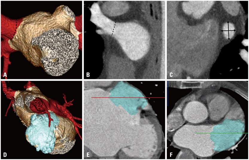
Fig. 1 Multiplanar reconstructed (MPR) images illustrating the double-oblique measurements of pulmonary veins (PVs) and left atrial appendage (LAA) ostial diameters. (A) 3D reconstruction image of left atrium and pulmonary veins. The dotted line denotes right superior PV ostium. (B) Oblique MPR view of the right superior PV. The ostium (dotted line) was confirmed in multiple views. (C) Enlarged axial MPR view across the ostium of the superior PV showing measurements of the PV diameter (black arrows). (D) 3D reconstruction image of LAA. (E and F) MPR view of the LAA showing enlarged LAA. 3D, three-dimensional.

Fig. 2 Comparison of LA volume (A) and orifice area of the LAA and PVs (B) between the three patient groups. LA, left atrium; LAA, left atrial appendage; LSPV, left superior pulmonary vein; LIPV, left inferior pulmonary vein; RSPV, right superior pulmonary vein; RIPV, right inferior pulmonary vein.

Fig. 3 ROC curve analysis. The LAA orifice area, LA volume, LIPV orifice area, and LSPV orifice area predicted stroke in AF patients with area under the curves (AUCs) of 0.739, 0.720, 0.674, and 0.587, respectively. ROC, receiver operating characteristic; LAA, left atrial appendage; LA, left atrium; LSPV, left superior pulmonary vein; LIPV, left inferior pulmonary vein; RSPV, right superior pulmonary vein; RIPV, right inferior pulmonary vein.
Reference
-
1. Cardiogenic brain embolism. The second report of the Cerebral Embolism Task Force. Arch Neurol. 1989; 46:727–743.2. Hart RG, Benavente O, McBride R, Pearce LA. Antithrombotic therapy to prevent stroke in patients with atrial fibrillation: a meta-analysis. Ann Intern Med. 1999; 131:492–501.
Article3. Furie KL, Kasner SE, Adams RJ, Albers GW, Bush RL, Fagan SC, et al. Guidelines for the prevention of stroke in patients with stroke or transient ischemic attack: a guideline for healthcare professionals from the american heart association/american stroke association. Stroke. 2011; 42:227–276.
Article4. Jaïs P, Haïssaguerre M, Shah DC, Chouairi S, Gencel L, Hocini M, et al. A focal source of atrial fibrillation treated by discrete radiofrequency ablation. Circulation. 1997; 95:572–576.
Article5. Haïssaguerre M, Jaïs P, Shah DC, Takahashi A, Hocini M, Quiniou G, et al. Spontaneous initiation of atrial fibrillation by ectopic beats originating in the pulmonary veins. N Engl J Med. 1998; 339:659–666.
Article6. Teh AW, Kalman JM, Lee G, Medi C, Heck PM, Ling LH, et al. Electroanatomic remodelling of the pulmonary veins associated with age. Europace. 2012; 14:46–51.
Article7. Casaclang-Verzosa G, Gersh BJ, Tsang TS. Structural and functional remodeling of the left atrium: clinical and therapeutic implications for atrial fibrillation. J Am Coll Cardiol. 2008; 51:1–11.8. Herweg B, Sichrovsky T, Polosajian L, Rozenshtein A, Steinberg JS. Hypertension and hypertensive heart disease are associated with increased ostial pulmonary vein diameter. J Cardiovasc Electrophysiol. 2005; 16:2–5.
Article9. Liu T, Li G. Pulmonary vein dilatation: another possible crosslink between left atrial enlargement and atrial fibrillation? Int J Cardiol. 2008; 123:193–194.
Article10. Beinart R, Heist EK, Newell JB, Holmvang G, Ruskin JN, Mansour M. Left atrial appendage dimensions predict the risk of stroke/TIA in patients with atrial fibrillation. J Cardiovasc Electrophysiol. 2011; 22:10–15.
Article11. Hunt SA, Abraham WT, Chin MH, Feldman AM, Francis GS, Ganiats TG, et al. 2009 Focused update incorporated into the ACC/AHA 2005 Guidelines for the Diagnosis and Management of Heart Failure in Adults A Report of the American College of Cardiology Foundation/American Heart Association Task Force on Practice Guidelines Developed in Collaboration With the International Society for Heart and Lung Transplantation. J Am Coll Cardiol. 2009; 53:e1–e90.12. European Heart Rhythm Association. European Association for Cardio-Thoracic Surgery. Camm AJ, Kirchhof P, Lip GY, Schotten U, et al. Guidelines for the management of atrial fibrillation: the Task Force for the Management of Atrial Fibrillation of the European Society of Cardiology (ESC). Eur Heart J. 2010; 31:2369–2429.13. Kim YH, Marom EM, Herndon JE 2nd, McAdams HP. Pulmonary vein diameter, cross-sectional area, and shape: CT analysis. Radiology. 2005; 235:43–49.
Article14. Yuan XP, Bach D, Skanes A, Drangova M. Assessment of intra- and interobserver variability of pulmonary vein measurements from CT angiography. Acad Radiol. 2004; 11:1211–1218.15. Maeda S, Iesaka Y, Otomo K, Uno K, Nagata Y, Suzuki K, et al. No severe pulmonary vein stenosis after extensive encircling pulmonary vein isolation: 12-month follow-up with 3D computed tomography. Heart Vessels. 2011; 26:440–448.
Article16. Lemola K, Sneider M, Desjardins B, Case I, Chugh A, Hall B, et al. Effects of left atrial ablation of atrial fibrillation on size of the left atrium and pulmonary veins. Heart Rhythm. 2004; 1:576–581.
Article17. Schwartzman D, Lacomis J, Wigginton WG. Characterization of left atrium and distal pulmonary vein morphology using multidimensional computed tomography. J Am Coll Cardiol. 2003; 41:1349–1357.
Article18. Manghat NE, Mathias HC, Kakani N, Hamilton MC, Morgan-Hughes G, Roobottom CA. Pulmonary venous evaluation using electrocardiogram-gated 64-detector row cardiac CT. Br J Radiol. 2012; 85:965–971.
Article19. Di Biase L, Santangeli P, Anselmino M, Mohanty P, Salvetti I, Gili S, et al. Does the left atrial appendage morphology correlate with the risk of stroke in patients with atrial fibrillation? Results from a multicenter study. J Am Coll Cardiol. 2012; 60:531–538.
Article20. Hur J, Kim YJ, Nam JE, Choe KO, Choi EY, Shim CY, et al. Thrombus in the left atrial appendage in stroke patients: detection with cardiac CT angiography--a preliminary report. Radiology. 2008; 249:81–87.
Article21. Benjamin EJ, D'Agostino RB, Belanger AJ, Wolf PA, Levy D. Left atrial size and the risk of stroke and death. The Framingham Heart Study. Circulation. 1995; 92:835–841.22. Black IW, Hopkins AP, Lee LC, Walsh WF. Evaluation of transesophageal echocardiography before cardioversion of atrial fibrillation and flutter in nonanticoagulated patients. Am Heart J. 1993; 126:375–381.
Article23. Fuster V, Rydén LE, Cannom DS, Crijns HJ, Curtis AB, Ellenbogen KA, et al. 2011 ACCF/AHA/HRS focused updates incorporated into the ACC/AHA/ESC 2006 guidelines for the management of patients with atrial fibrillation: a report of the American College of Cardiology Foundation/American Heart Association Task Force on practice guidelines. Circulation. 2011; 123:e269–e367.24. Goldstein LB, Adams R, Alberts MJ, Appel LJ, Brass LM, Bushnell CD, et al. Primary prevention of ischemic stroke: a guideline from the American Heart Association/American Stroke Association Stroke Council: cosponsored by the Atherosclerotic Peripheral Vascular Disease Interdisciplinary Working Group; Cardiovascular Nursing Council; Clinical Cardiology Council; Nutrition, Physical Activity, and Metabolism Council; and the Quality of Care and Outcomes Research Interdisciplinary Working Group. Circulation. 2006; 113:e873–e923.25. Kim YL, Joung B, On YK, Shim CY, Lee MH, Kim YH, et al. Early experience using a left atrial appendage occlusion device in patients with atrial fibrillation. Yonsei Med J. 2012; 53:83–90.
Article26. Wang Y, Di Biase L, Horton RP, Nguyen T, Morhanty P, Natale A. Left atrial appendage studied by computed tomography to help planning for appendage closure device placement. J Cardiovasc Electrophysiol. 2010; 21:973–982.
Article27. Douglas YL, Jongbloed MR, Gittenberger-de Groot AC, Evers D, Dion RA, Voigt P, et al. Histology of vascular myocardial wall of left atrial body after pulmonary venous incorporation. Am J Cardiol. 2006; 97:662–670.
Article28. Shirani J, Alaeddini J. Structural remodeling of the left atrial appendage in patients with chronic non-valvular atrial fibrillation: implications for thrombus formation, systemic embolism, and assessment by transesophageal echocardiography. Cardiovasc Pathol. 2000; 9:95–101.
Article29. Mansour M, Mandapati R, Berenfeld O, Chen J, Samie FH, Jalife J. Left-to-right gradient of atrial frequencies during acute atrial fibrillation in the isolated sheep heart. Circulation. 2001; 103:2631–2636.
Article30. Hart RG, Halperin JL. Atrial fibrillation and stroke: concepts and controversies. Stroke. 2001; 32:803–808.31. Hassink RJ, Aretz HT, Ruskin J, Keane D. Morphology of atrial myocardium in human pulmonary veins: a postmortem analysis in patients with and without atrial fibrillation. J Am Coll Cardiol. 2003; 42:1108–1114.32. Tsao HM, Yu WC, Cheng HC, Wu MH, Tai CT, Lin WS, et al. Pulmonary vein dilation in patients with atrial fibrillation: detection by magnetic resonance imaging. J Cardiovasc Electrophysiol. 2001; 12:809–813.
Article33. Lin WS, Prakash VS, Tai CT, Hsieh MH, Tsai CF, Yu WC, et al. Pulmonary vein morphology in patients with paroxysmal atrial fibrillation initiated by ectopic beats originating from the pulmonary veins: implications for catheter ablation. Circulation. 2000; 101:1274–1281.
Article34. Kato R, Lickfett L, Meininger G, Dickfeld T, Wu R, Juang G, et al. Pulmonary vein anatomy in patients undergoing catheter ablation of atrial fibrillation: lessons learned by use of magnetic resonance imaging. Circulation. 2003; 107:2004–2010.
Article
- Full Text Links
-
- Actions
-
Cited
- CITED
-
- Close
- Share
-
- Similar articles
-
- Controlled Atrial Fibrillation after Pulmonary Vein Stenting
- Practical Issues to Prevent Stroke Associated with Non-valvular Atrial Fibrillation
- The Mechanism of and Preventive Therapy for Stroke in Patients with Atrial Fibrillation
- Cardioembolic Stroke in Atrial Fibrillation-Rationale for Preventive Closure of the Left Atrial Appendage
- The Joint Multicenter Study on the Atrial Fibrillation in Korea
