J Korean Soc Spine Surg.
2015 Sep;22(3):123-126. 10.4184/jkss.2015.22.3.123.
Surgical Treatment of Spinal Extradural Arachnoid Cyst: A Case Report
- Affiliations
-
- 1Department of Orthopedic Surgery, Eulji University School of Medicine, Daejeon, Korea. hjkim@eulji.ac.kr
- KMID: 2068932
- DOI: http://doi.org/10.4184/jkss.2015.22.3.123
Abstract
- STUDY DESIGN: A case report.
OBJECTIVES
To report a case of spinal extradural arachnoid cyst. SUMMARY OF LITERATURE REVIEW: Extradural arachonid cysts of the spine are a rare cause of spinal cord and nerve root compression. There are few reports about it, and the etiology remains unclear.
MATERIALS AND METHODS
The authors performed a clinical and radiographic case review.
RESULTS
A 56-year-old male patient presented with both lower extremity radiating pain and tingling sensation in both feet for four years. His MRI revealed a large, well-demarcated extradural lesion, isointense to cerebrospinal fluid from L1 to L3. We performed dural repair and laminectomy for partial resection of the cyst. The outcome was good in the immediate postoperative period, and the patient made a full recovery without complications.
CONCLUSIONS
Surgical treatment should be considered for large spinal extradural arachnoid cysts with neurologic symptoms when conservative treatment does not work.
MeSH Terms
Figure
-

Fig. 1. A preoperative MRI shows an arachnoid cyst from L1 to L3. A Hypointense T1-weighted image (A) Hyperintense T2-weighted image (B) An axial view (C, D) Shows dural compression.

Fig. 2. A lateral view of the myelogram. There is no communication between the cyst and subarachnoid space.
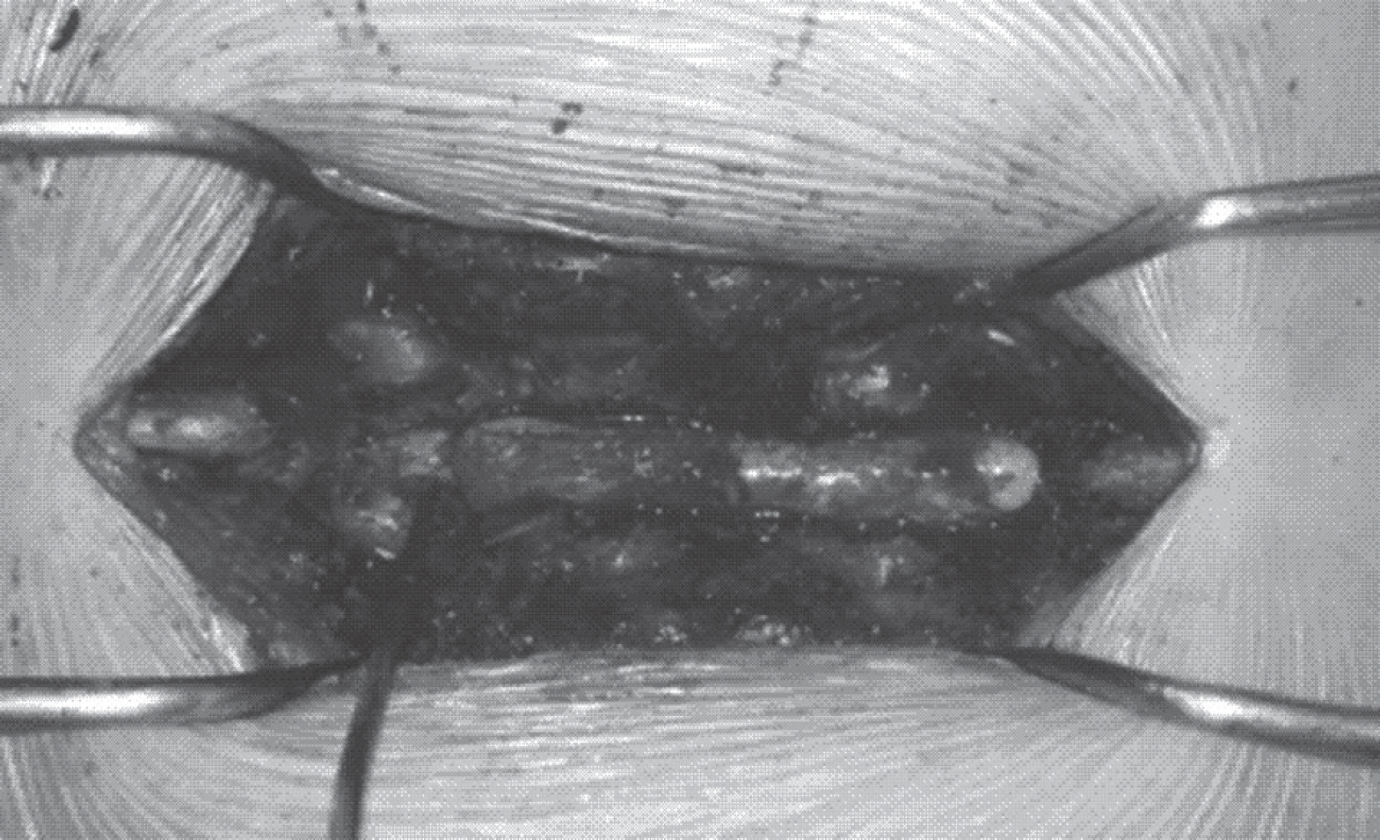
Fig. 3. An intraoperative view showing a grayish translucent cyst.
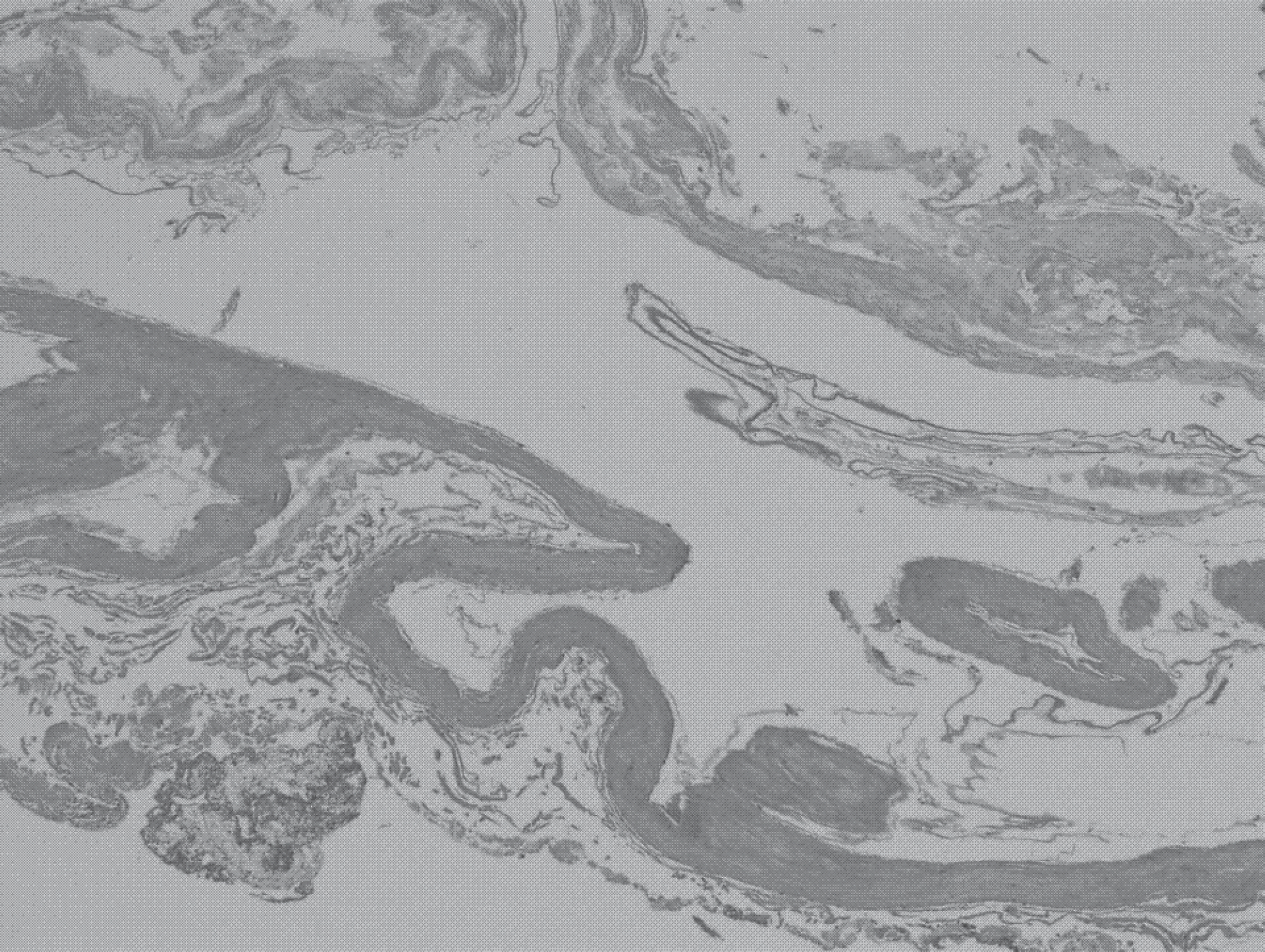
Fig. 4. A histopathological examination of the cyst wall showed a fibro-cartilagenous layer with a thin layer of simple squamous epithelium.
Reference
-
1. Fortuna A, La Torre E, Ciappetta P. Arachnoid diverticula: a unitary approach to spinal cysts communicating with the subarachnoid space. Acta Neurochir. 1977; 39:259–68.
Article2. Cilluffo JM, Gomez MR, Reese DF, et al. �diopathic (�congenital”) spinal arachnoid diverticula. Clinical diagnosis and surgical results. Myo Clin Proc. 1998; 56:93–101.3. Fortuna A, La Torre E, Ciappetta P. Arachnoidal diverticula: a unitary approach to spinal cysts communicating with the subarachnoid space. Acta Neurochir. 1977; 39:259–68.4. Nabors MW, Pait TG, Byrd EB, et al. Updated assessment and current classification of spinal meningeal cysts. J Neurosurg. 1988; 68:366–77.
Article5. Bergland RM. Congenital intraspinal extradural cyst. Report of three cases in one family. J Neurosug. 1968; 28:495–9.6. Liu JK, Cole CD, Kan P, et al. Spinal extradural arachnoid cysts: clinical, radiological, and surgical features. Neurosurg Focus. 2007; 22:E6.
Article7. Ersahin Y, Yildizhan A, Seber N. Spinal extradural arachnoid cyst. Childs Nerv Syst. 1993; 9:250–2.
Article8. Lee HJ, Cho WH, Han � H, et al. Large thoracolumbar extradural arachnoid cyst excised by minimal skipped hemilaminectomy: A case report. Korean J spine. 2013; 10:28–31.
Article9. Neo M, Koyoma T, Sakamoto T, et al. Detection of a dural defect by cinematic magnetic resonance imaging and its se-lective closure as a treatment for a spinal extradural arachnoid cyst. Spine (Phila Pa 1976). 2004; 29:E426–30.
Article10. Stechison MT, Hendrick EB, Cohen E. Spinal extradural arachnoid cyst. Pedatr Neurosci. 1989; 15:36–8.
Article
- Full Text Links
-
- Actions
-
Cited
- CITED
-
- Close
- Share
-
- Similar articles
-
- Multiple Extradural Arachnoid Cyst : A Case Report
- Extradural Spinal Arachnoid Cyst as a Cause of Cauda Equina Syndrome in a Child
- Spinal Extradural Arachnoid Cyst: Minimally Invasive Surgical Treatment after Localization of Dural Defect Using Magnetic Resonance Myelogram
- Septated Extradural Arachnoid Cyst in Thoracolumbar Spine Causing Myelopathy
- Spinal Extradural Arachnoid Cyst: Case Report
