Leptomeningeal Carcinomatosis of Gastric Cancer Misdiagnosed as Vestibular Schwannoma
- Affiliations
-
- 1Department of Neurosurgery, Chung-Ang University College of Medicine, Seoul, Korea. jtkwon@cau.ac.kr
- 2Department of Otorhinolaryngology-Head and Neck Surgery, Chung-Ang University College of Medicine, Seoul, Korea.
- KMID: 2067077
- DOI: http://doi.org/10.3340/jkns.2014.56.1.51
Abstract
- Gastric cancer is one of the most common causes of cancer-related death in Asian countries, including Korea. We experienced a case of leptomeningeal carcinomatosis (LC) from gastric cancer that was originally misdiagnosed as vestibular schwannoma based on the similar radiological characteristics. To our knowledge, LC from gastric cancer is very rare. In conclusion, our experience with this case suggests that clinicians should consider the possibility of delayed leptomeningeal metastasis when treating patients with gastric cancer.
Keyword
MeSH Terms
Figure
-

Fig. 1 On T1-weighted axial image (A) and a 3-dimensional fluid attenuated inversion recovery VISTA CE one (B), taken at the patient's initial visit due to hearing difficulty, there are multiple enhanced lesions along the cisternal and canalicular segments of the bilateral vestibulocochlear nerves.
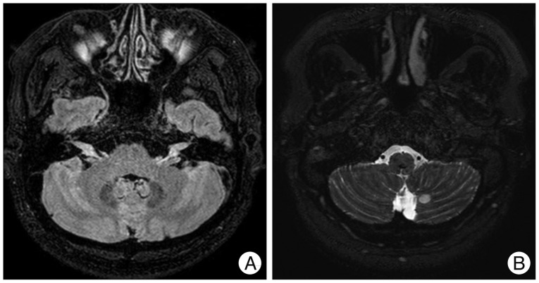
Fig. 2 On a 3-dimensional fluid attenuated inversion recovery VISTA CE image (A), there is an interval growth of enhanced areas along the cisternal and canalicular segments of the bilateral eighth nerves and the newly developed intense enhancement of the bilateral trigeminal nerves involving the cisternal segments and Meckel's caves. In addition, on a 3-dimensional DRIVE axial image (B), there is a newly developed, circular enhancing area in the left medial cerebellum.

Fig. 3 The enhanced magnetic resonance image of the spine. On T1-weighted turbo spin-echo sagittal image (A) and axial image (B), there are multifocal enhanced lesions in the conus medullaris and cauda equine. These findings are suggestive of metastatic tumors.

Fig. 4 Positron emission tomography-computed tomography. On transverse image (A) of low-dose head CT, there are non-hypermetabolic multiple brain nodules. These findings are suggestive of benign lesion. In addition on torso maximal intensity projection coronal view image (B) of the whole body, there are no metastatic lesions in any other sites.

Fig. 5 An intraoperative microscopic image shows a whitish, mucoid-like mass of 1 cm in diameter around the vestibulocochlear nerve.
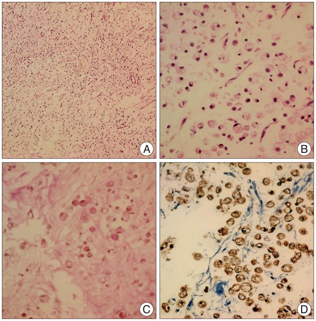
Fig. 6 A histopathologic image of the brain tumor shows discohesive, pleomorphic, malignant cells with a signet-ring appearance, with a prominent intracytoplasmic mucin vacuole where the nucleus is peripherally dispersed. (A) Hematoxylin and eosin stain, ×100; (B) hematoxylin and eosin stain, ×200; (C) mucicarmine stain (+), ×200; and (D) CK stain (+), ×200.
Reference
-
1. Bruna J, González L, Miró J, Velasco R, Gil M, Tortosa A, et al. Leptomeningeal carcinomatosis : prognostic implications of clinical and cerebrospinal fluid features. Cancer. 2009; 115:381–389. PMID: 19109820.2. Bulut G, Erden A, Karaca B, Göker E. Leptomeningeal carcinomatosis of gastric adenocarcinoma. Turk J Gastroenterol. 2011; 22:195–198. PMID: 21796558.
Article3. Chamberlain MC. Leptomeningeal metastasis. Semin Neurol. 2010; 30:236–244. PMID: 20577930.
Article4. Groves MD. New strategies in the management of leptomeningeal metastases. Arch Neurol. 2010; 67:305–312. PMID: 20212228.
Article5. Larson DA, Rubenstein JL, Mcdermott MW. Treatment of metastatic cancer. In : DeVita JVT, Hellman S, Rosenberg SA, editors. Cancer : Principles and Practice of Oncology. ed 7. Philadelphia: Lippincott Williams & Wilkins;2005. p. 2333.6. Lisenko Y, Kumar AJ, Yao J, Ajani J, Ho L. Leptomeningeal carcinomatosis originating from gastric cancer : report of eight cases and review of the literature. Am J Clin Oncol. 2003; 26:165–170. PMID: 12714889.7. Oh SY, Lee SJ, Lee J, Lee S, Kim SH, Kwon HC, et al. Gastric leptomeningeal carcinomatosis : multi-center retrospective analysis of 54 cases. World J Gastroenterol. 2009; 15:5086–5090. PMID: 19860003.
Article8. Park KK, Yang SI, Seo KW, Kim YO, Yoon KY. A case of metastatic leptomeningeal carcinomatosis from early gastric carcinoma. World J Surg Oncol. 2012; 10:74. PMID: 22553956.
Article9. Pentheroudakis G, Pavlidis N. Management of leptomeningeal malignancy. Expert Opin Pharmacother. 2005; 6:1115–1125. PMID: 15957966.
Article10. Sandberg DI, Bilsky MH, Souweidane MM, Bzdil J, Gutin PH. Ommaya reservoirs for the treatment of leptomeningeal metastases. Neurosurgery. 2000; 47:49–54. discussion 54-55. PMID: 10917346.
Article11. Shin HR, Jung KW, Won YJ, Kong HJ, Yim SH, Sung J, et al. National cancer incidence for the year 2002 in Korea. Cancer Res Treat. 2007; 39:139–149. PMID: 19746208.
Article12. Tomita H, Yasui H, Boku N, Nakasu Y, Mitsuya K, Onozawa Y, et al. Leptomeningeal carcinomatosis associated with gastric cancer. Int J Clin Oncol. 2012; 17:361–366. PMID: 21847535.
Article
- Full Text Links
-
- Actions
-
Cited
- CITED
-
- Close
- Share
-
- Similar articles
-
- A Case of Leptomeningeal Carcinomatosis Presenting as a Neurological Complication of Stomach Cancer
- Intrathecal Trastuzumab Treatment in Patients with Breast Cancer and Leptomeningeal Carcinomatosis
- A Case of Gastric Adenocarcinoma Presenting as Meningeal Carcinomatosis
- Hypervascular Vestibular Schwannoma: A Case Report
- Diagnosis and Management of Vestibular Schwannoma: Focus on Dizziness
