J Korean Endocr Soc.
2008 Apr;23(2):137-141. 10.3803/jkes.2008.23.2.137.
Non-functional Pituitary Adenoma Detected on (18)F-fluorodeoxyglucose Positron Emission Tomography ((18)F-FDG-PET) in a Patient with Mucosa-associated Lymphoid Tissue Lymphoma
- Affiliations
-
- 1Department of Internal Medicine, Yonsei University College of Medicine, Seoul, Korea.
- 2Endocrine Research Institute, Yonsei University College of Medicine, Seoul, Korea.
- 3Department of Nuclear Medicine, Yonsei University College of Medicine, Seoul, Korea.
- KMID: 2063565
- DOI: http://doi.org/10.3803/jkes.2008.23.2.137
Abstract
- Magnetic resonance imaging (MRI) is the modality of choice for the detection and characterization of a pituitary adenoma. Uptake of (18)F-fluorodeoxyglucose (FDG) by intrasellar tumors, including pituitary adenomas, has been reported in several previous studies. We report a case where a pituitary adenoma was detected on FDG-positron emission tomography (PET), but the tumor was not detected with the use of sellar MRI. A 31-year-old woman was referred to the clinic due to a focal increase of FDG uptake at the pituitary fossa seen on whole body FDG-PET. The patient was receiving chemotherapy due to a recurred B-cell lymphoma of the mucosa-associated lymphoid tissue type. Subsequently, sellar MRI was performed, and images showed a small non-enhancing heterogenous cystic lesion in the midline of the pituitary gland, radiologically suggestive of a Rathke's cleft cyst. However, sellar MRI failed to identify a lesion consistent with a pituitary tumor that corresponded to the site of increased FDG uptake detected by the use of PET, despite the inclusion of a dynamic contrast enhanced sequence. Despite the negative findings of the MRI examination, basal and stimulated levels of the GnRH free alpha-subunit were profoundly increased. Therefore, we suspected the presence of a non-functional pituitary tumor in addition to a Rathke's cleft cyst, rather than pituitary involvement of a lymphoma, based on the hormone levels and PET scan findings.
Keyword
MeSH Terms
Figure
-

Fig. 1 Midline sagittal section and coronal section of 18F-FDG-PET. Focal FDG uptake at the pituitary gland (arrow).

Fig. 2 GnRH and TRH stimulation test. The basal and GnRH stimulated levels of free α-subunit were increased more than three times the normal range. TRH stimulation test showed no significant change.
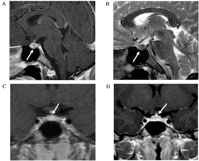
Fig. 3 Midline sagittal image of gadolinium-enhanced T1 (A), T2 (B) weighted MRI and coronal image of dynamic (C),. gadolinium-enhanced T1 (D). A small non-enhancing nodule in the middle portion of pituitary gland, suggestive of Rathke's cleft cyst (arrow).
Reference
-
1. Janine R, Arnd D. Imaging of sellar and parasellar lesions. Clin Neurol Neurosurg. 2007. 109:111–112.2. Lee K, Lee E, Kim K, Chung Y, Lee B, Park S, Lim S, Lee H, Yoon D, Kim Y, Huh K. Alpha-subunit secretion of pituitary adenomas. J Kor Endocrinol. 1993. 8:127–133.3. Lee E, Kin K, Lee H, Nam M, Jo J, Song Y, Lim S, Jung H, Kim T, Kim S, Choi J, Chung S, Lee K, Huh K. Endocrinological and morphological characteristics of clinically nonfunctioning pituitary adenoma. J Kor Endocrinol. 1994. 9:200–202.4. Richard J, Odiled D, Adam M. Pituitary adenoma detected on FDG positron emission tomography in a patient with mucosa-associated lymphoid tissue lymphoma. Clin Nucl Med. 2003. 28:296–298.5. Tsuyoshi K, William H, Alan L, Dominique D. Serendipitous detection of Cushing's disease by FDG positron emission tomography and a review of the literature. Clin Nucl Med. 2002. 27:176–178.6. Mats B, Carin M, Lundberg P, Bengt L. PET as a tool in the clinical evaluation of pituitary adenomas. J Nucl Med. 1991. 32:610–615.7. Zoran R. Pituitary adenomas. Top Magn Reson Imaging. 2005. 16:277–288.8. Guy RL, Benn JJ, Ayers AB, Bingham JB, Lowy C, Cox TC, Sonksen PH. A comparison of CT and MRI on the assessment of the pituitary and paraseller region. Clin Radiol. 1991. 43:156–161.9. Stein AL, Levenick MN, Kletzky OA. Computed tomography versus magnetic resonance imaging for the evaluation of suspected pituitary adenomas. Obstet Gynecol. 1989. 73:996–999.10. Carin M. Positron emission tomography in acromegaly and other pituitary adenoma patients. Neuroendocrinology. 2006. 83:205–210.11. Chanson P, Brochier S. Non-functioning pituitary adenomas. J Endocrinol Invest. 2005. 28:93–99.12. De Souza B, Brunetti A, Fulham MJ, Brooks RA, DeMichele D, Cook P, Nieman L, Doppman JL, Oldfield EH, Di Chiro G. Pituitary microadenomas: a PET study. Radiology. 1990. 177:39–44.13. Francavilla TL, Miletich RS, DeMichele D. Position emission tomography of pituitary macroadenoma: hormone production and effects of therapies. Neurosurgery. 1991. 28:826–833.14. Wong CY, Thie J, Parling-Lynch KJ, Zakalik D, Wong RH, Gaskill M, Margolis JH, Hill J, Sukari A, Chundru S, Fink-Bennett D, Nagle C. Investigating the existence of quantum metabolic values in non-Hodgkin's lymphoma by 2-deoxy-2-[F-18]fluoro-D-glucose positron emission tomography. Mol Imaging Biol. 2007. 9:43–49.15. Megan Ogilvie C, Payne S, Evanson J, Lister TA, Grossman AB. Lymphoma metastasizing to the pituitary: an unusual presentation of a treatable disease. Pituitary. 2005. 8:139–146.16. Sumida M, Arita K, Migita K, Tomonaga A, Iida K, Kurisu K. Concomitant pituitary adenoma and Rathke's cleft cyst. Neuroendocrinology. 2001. 43:755–759.
- Full Text Links
-
- Actions
-
Cited
- CITED
-
- Close
- Share
-
- Similar articles
-
- A non-functioning pituitary macroadenoma detected incidentally with PET-CT
- Unexpected Second Primary Malignancies Detected by F-18 FDG PET/CT During Follow-up for Primary Malignancy: Two Case Reports
- Efficacy of Positron Emission Tomography/Computed Tomography in Gastric Mucosa-associated Lymphoid Tissue Lymphoma
- F-18 fluorodeoxyglucose positron emission tomography/computed tomography in the infection of heart
- Whole Body Positron Emission Tomography/Computed Tomography
