Hip Pelvis.
2014 Jun;26(2):115-123. 10.5371/hp.2014.26.2.115.
Correlation of Structural Bony Abnormalities and Mechanical Symptoms of Hip Joints
- Affiliations
-
- 1Department of Orthopaedic Surgery, Seoul National University Bundang Hospital, Seoul National University College of Medicine, Seoul, Korea.
- 2Department of Orthopaedic Surgery, Chung-Ang University College of Medicine, Seoul, Korea. hayongch@naver.com
- KMID: 2054173
- DOI: http://doi.org/10.5371/hp.2014.26.2.115
Abstract
- PURPOSE
The purpose of this study is to determine structural bony abnormalities predisposing for femoroacetabular impingement by comparison of patients with and without mechanical symptoms.
MATERIALS AND METHODS
We conducted this comparative study on 151 patients (151 hips; mean age 44.8 years; range 16-73 years) with mechanical symptoms with results of multi-detector computed tomography (MDCT) arthrography (the symptomatic group). Each patient was matched with a control who underwent MDCT due to ureter stone (the asymptomatic group) in terms of age, gender, site (right or left), and time at diagnosis. Acetabular evaluations, which included cranial and central anteversion and anterior and lateral center edge angles and femoral measurements, were performed. In addition, we evaluated the prevalence and characteristics of structural bone abnormalities between the two groups.
RESULTS
The prevalence for patients who had at least one structural bony abnormality in the symptomatic and asymptomatic groups was 80.1% (121/151) and 54.3% (82/151), respectively (odds ratio: 3.39, 95% confidence interval: 2.30-5.66; P<0.001). The most common osseous abnormality was the isolated Pincer type in both groups: 89 (73.6%) of 121 hips with an osseous abnormality in the symptomatic group and 57 (69.5%) of 82 hips with an osseous abnormality in the asymptomatic group. By analysis of CT arthrography in symptomatic patients, a labral tear was found in 107 hips (70.9%), and 86 (80%) of these hips had a structural bony abnormality.
CONCLUSION
A significantly greater prevalence rate of structural bony abnormality was observed for the symptomatic group than for the asymptomatic group. These findings are helpful for development of appropriate treatment plans.
MeSH Terms
Figure
-
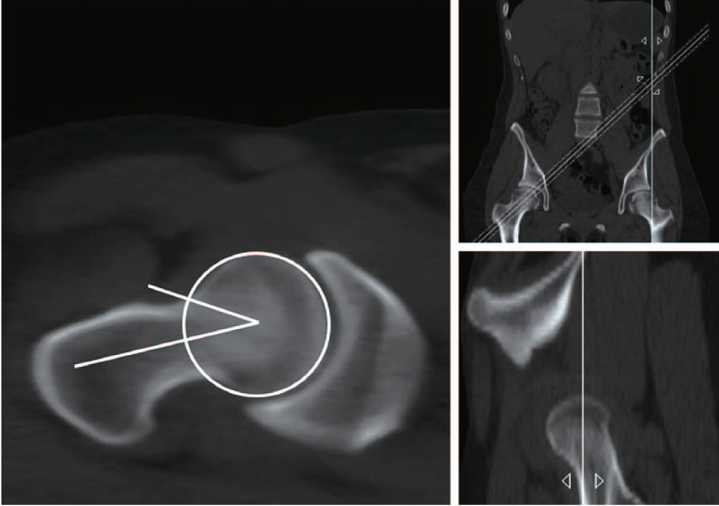
Fig. 1 Alpha angle measurement with reformatted pelvis multi-detector computed tomography scan.
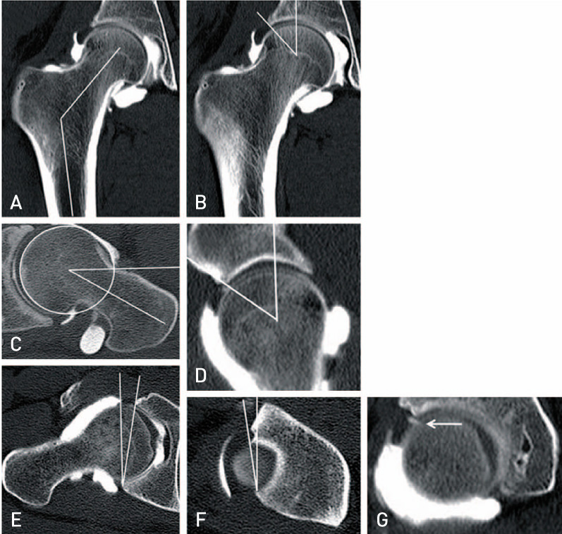
Fig. 2 Assessment parameters with using multi-detector computed tomography arthrography. (A) Neck shaft angle, (B) lateral center edge angle, (C) alpha angle, (D) anterior center edge angle, (E) central acetabular version, (F) cranial acetabular version, and (G) labral abnormality (arrow).
Reference
-
1. Kang AC, Gooding AJ, Coates MH, Goh TD, Armour P, Rietveld J. Computed tomography assessment of hip joints in asymptomatic individuals in relation to femoroacetabular impingement. Am J Sports Med. 2010; 38:1160–1165.
Article2. Beck M, Kalhor M, Leunig M, Ganz R. Hip morphology influences the pattern of damage to the acetabular cartilage: femoroacetabular impingement as a cause of early osteoarthritis of the hip. J Bone Joint Surg Br. 2005; 87:1012–1018.3. Arai N, Nakamura S, Matsushita T. Difference between 2 measurement methods of version angles of the acetabular component. J Arthroplasty. 2007; 22:715–720.
Article4. Hassan DM, Johnston GH, Dust WN, Watson LG, Cassidy D. Radiographic calculation of anteversion in acetabular prostheses. J Arthroplasty. 1995; 10:369–372.
Article5. Widmer KH. A simplified method to determine acetabular cup anteversion from plain radiographs. J Arthroplasty. 2004; 19:387–390.
Article6. Clohisy JC, Carlisle JC, Trousdale R, et al. Radiographic evaluation of the hip has limited reliability. Clin Orthop Relat Res. 2009; 467:666–675.
Article7. Choi JY, Kang HS, Hong SH, et al. Optimization of the contrast mixture ratio for simultaneous direct MR and CT arthrography: an in vitro study. Korean J Radiol. 2008; 9:520–525.
Article8. Farber JM. CT arthrography and postoperative musculoskeletal imaging with multichannel computed tomography. Semin Musculoskelet Radiol. 2004; 8:157–166.
Article9. Wenger DE, Kendell KR, Miner MR, Trousdale RT. Acetabular labral tears rarely occur in the absence of bony abnormalities. Clin Orthop Relat Res. 2004; 145–150.
Article10. Peelle MW, DellaRocca GJ, Maloney WJ, Curry MC, Clohisy JC. Acetabular and femoral radiographic abnormalities associated with labral tears. Clin Orthop Relat Res. 2005; 441:327–333.
Article11. Tonnis D. Normal values of the hip joint for the evaluation of X-rays in children and adults. Clin Orthop Relat Res. 1976; 39–47.12. Wiberg G. Studies on dysplastic acetabula and congenital subluxation of the hip joint. Acta Chir Scand. 1939; 83:1–135.13. Tonnis D, Heinecke A. Acetabular and femoral anteversion: relationship with osteoarthritis of the hip. J Bone Joint Surg Am. 1999; 81:1747–1770.14. Lequesne M, Seze S. Le faux profil du bassin. Nouvelle incidence radiographique pour l'etude de la hanche. Son utilite dans les dysplasies et les differentes coxopathies. Rev Rhum Mal Osteoartic. 1961; 28:643–652.15. Reynolds D, Lucas J, Klaue K. Retroversion of the acetabulum. A cause of hip pain. J Bone Joint Surg Br. 1999; 81:281–288.16. Notzli HP, Wyss TF, Stoecklin CH, Schmid MR, Treiber K, Hodler J. The contour of the femoral head-neck junction as a predictor for the risk of anterior impingement. J Bone Joint Surg Br. 2002; 84:556–560.17. Panzer S, Augat P, Esch U. CT assessment of herniation pits: prevalence, characteristics, and potential association with morphological predictors of femoroacetabular impingement. Eur Radiol. 2008; 18:1869–1875.
Article18. Montgomery AA, Graham A, Evans PH, Fahey T. Interrater agreement in the scoring of abstracts submitted to a primary care research conference. BMC Health Serv Res. 2002; 2:8.
Article19. Landis JR, Koch GG. The measurement of observer agreement for categorical data. Biometrics. 1977; 33:159–174.
Article20. Weir A, de Vos RJ, Moen M, Holmich P, Tol J. Prevalence of radiological signs of femoroacetabular impingement in patients presenting with long-standing adductor-related groin pain. Br J Sports Med. 2011; 45:6–9.
Article21. Beaule PE, Zaragoza E, Motamedi K, Copelan N, Dorey FJ. Three-dimensional computed tomography of the hip in the assessment of femoroacetabular impingement. J Orthop Res. 2005; 23:1286–1292.
Article22. Karachalios T, Karantanas AH, Malizos K. Hip osteoarthritis: what the radiologist wants to know. Eur J Radiol. 2007; 63:36–48.
Article23. Neumann G, Mendicuti AD, Zou KH, et al. Prevalence of labral tears and cartilage loss in patients with mechanical symptoms of the hip: evaluation using MR arthrography. Osteoarthritis Cartilage. 2007; 15:909–917.
Article24. Nishii T, Tanaka H, Sugano N, Miki H, Takao M, Yoshikawa H. Disorders of acetabular labrum and articular cartilage in hip dysplasia: evaluation using isotropic high-resolutional CT arthrography with sequential radial reformation. Osteoarthritis Cartilage. 2007; 15:251–257.
Article25. Yamamoto Y, Tonotsuka H, Ueda T, Hamada Y. Usefulness of radial contrast-enhanced computed tomography for the diagnosis of acetabular labrum injury. Arthroscopy. 2007; 23:1290–1294.
Article
- Full Text Links
-
- Actions
-
Cited
- CITED
-
- Close
- Share
-
- Similar articles
-
- Osteoarthritis of Hip in a Hypogammaglobulinemia Patient
- Comprehensive Review of Advancements in Hip Arthroscopy
- The Role of IVIRI in Early Ankylosing Spondylitis: Emphasis on the Sacroiliac and Hip Joints
- Osteotomy around the Hip Joint
- Changes of the hip joints associated with chronic subluxation and dislocation: CT and plain radiographic analysis
