Long-term Outcomes of Drug-eluting Stents in Symptomatic Intracranial Stenosis
- Affiliations
-
- 1Departments of Radiology and Research Institute of Radiology, University of Ulsan, College of Medicine, Asan Medical Center, Seoul, Korea. dcsuh@amc.seoul.kr
- KMID: 2052075
- DOI: http://doi.org/10.5469/neuroint.2013.8.1.9
Abstract
- PURPOSE
The use of drug-eluting stent (DES) to treat intracranial stenosis has shown short-term success. However, there are no reports regarding the long-term results of DES. We present the long-term clinical outcome after DES stenting for symptomatic severe intracranial stenosis.
MATERIALS AND METHODS
Our study included a consecutive series of 11 patients who underwent intracranial stenting using DES between March and July, 2006, during the time when bare metal stents were not available at our medical institution. The mean patient age was 59 years. Lesion location was the middle cerebral artery in five patients, the intradural vertebral artery in three, the basilar artery in one, the vertebrobasilar junction in one, and the cavernous internal cerebral artery in one patient. We evaluated the technical success, defined as reduction of residual stenosis < or =30% in the target lesion) as well as the clinical and imaging outcomes as long as 75 months following the procedure. In addition to a cerebral angiogram (n = 2), follow-up study was obtained by CT angiography (n = 6) or intracranial Doppler imaging (n = 2) during a mean time of 55 months after the procedure (range, 24 to 73 months). Three patients refused imaging follow-up and accepted only clinical follow-up. The mean clinical follow-up period was 67 months (range, 47-75 months).
RESULTS
Stenting in all patients was technically successful and without periprocedural complications. There was thrombus formation during the procedure in one patient who experienced no further complications. There were no new neurological events during the mean follow-up period of 5.6 years. No patients were found to have restenosis > or =50% at during the mean follow-up period of 55 months. One patient died of a sudden heart attack 59 months following the procedure which was regarded as unrelated to the cerebral lesion.
CONCLUSION
Our study demonstrates that DES shows long-term stability and safety, and results in good clinical outcomes with a low rate of restenosis.
MeSH Terms
Figure
-
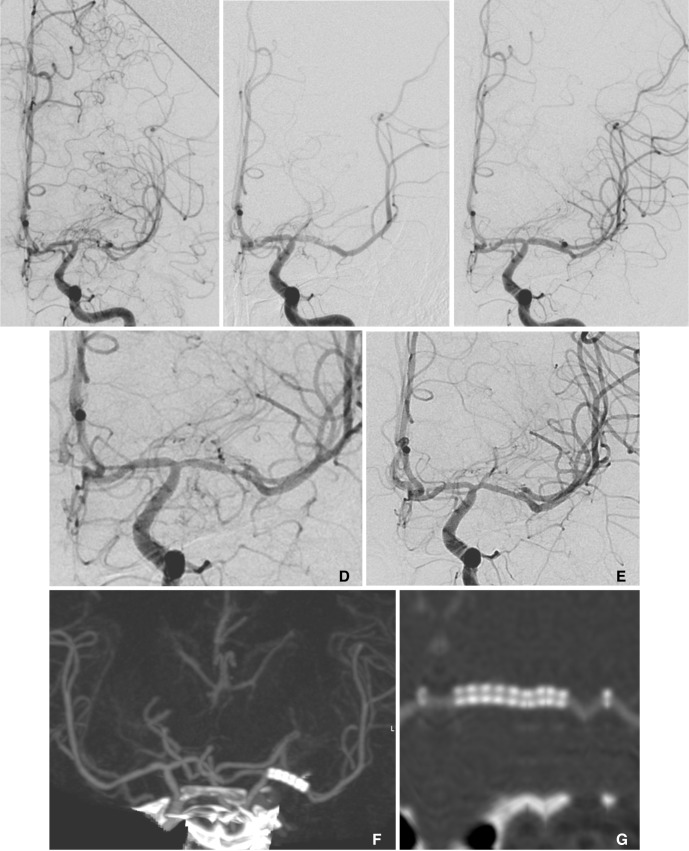
Fig. 1 A 35-year-old male presented with repeated TIAs against medication for 18 months. (A) The initial angiogram showed severe segmental stenosis of the left M1 and moyamoya-like collaterals around the stenosis. (B) There was an intraluminal filling defect after angioplasty and stenting, probably caused by incomplete antiplatelet medication or antiplatelet resistance. (C) The final angiogram obtained after intravenous antiplatelet agent infusion showed good patency of the M1. Also note the immediate disappearance of the moyamoya-like collaterals after stenting. (D) There was approximately 50% restenosis at the distal in-stent portion of the left M1, as seen on the 13-month follow-up angiogram, although the patient was without any symptoms. Cilostazol was added-added to the medication. (E) The restenosis had disappeared on the angiogram obtained 25 months later. (F) There was no symptom recurrence or new symptoms and no evidence of restenosis on the CT angiography obtained 41 months later. Note the symmetric filling of both MCA branches. (G) The linearly reconstructed stent lumen shows luminal patency. Transcranial Doppler examination at 73 months showed slightly increased but similar peak systolic velocities in both M1s.
Cited by 3 articles
-
Long-term outcomes of drug-eluting stent implantation in patients with symptomatic extra- and intracranial atherosclerotic stenoses
Junhyung Kim, Seung Pil Ban, Young Deok Kim, O-Ki Kwon
J Cerebrovasc Endovasc Neurosurg. 2020;22(4):216-224. doi: 10.7461/jcen.2020.E2020.09.001.Comparison of Drug-eluting Coronary Stents, Bare Coronary Stents and Self-expanding Stents in Angioplasty of Middle Cerebral Artery Stenoses
Jong-Hyeog Lee, Sung-Min Jo, Kwang-Deog Jo, Moon-Kyu Kim, Sang-Youl Lee, Seung-Hoon You
J Cerebrovasc Endovasc Neurosurg. 2013;15(2):85-95. doi: 10.7461/jcen.2013.15.2.85.Neurotoxicity of Paclitaxel and Rapamycin in a Rat Model with Transient Blood-Brain Barrier Opening
Won-Sang Cho, Jung Hoon Choi, O-Ki Kwon
J Korean Neurosurg Soc. 2022;65(2):180-185. doi: 10.3340/jkns.2021.0077.
Reference
-
1. Kasner SE, Chimowitz MI, Lynn MJ, Howlett-Smith H, Stern BJ, Hertzberg VS, et al. Predictors of ischemic stroke in the territory of a symptomatic intracranial arterial stenosis. Circulation. 2006; 113:555–563. PMID: 16432056.
Article2. Thijs VN, Albers GW. Symptomatic intracranial atherosclerosis: outcome of patients who fail antithrombotic therapy. Neurology. 2000; 55:490–497. PMID: 10953179.
Article3. Suh DC, Ko YB, Park ST, Yoon K, Lim OK, Oh JS, et al. Computational flow dynamics of the severe m1 stenosis before and after stenting. Neurointervention. 2011; 6:13–16. PMID: 22125742.
Article4. Lu PH, Park JW, Park S, Kim JL, Lee DH, Kwon SU, et al. Intracranial stenting of subacute symptomatic atherosclerotic occlusion versus stenosis. Stroke. 2011; 42:3470–3476. PMID: 21940974.
Article5. SSYLVIA study investigators. Stenting of symptomatic atherosclerotic lesions in the vertebral or intracranial arteries (SSYLVIA): study results. Stroke. 2004; 35:1388–1392. PMID: 15105508.6. Qureshi AI, Kirmani JF, Hussein HM, Harris-Lane P, Divani AA, Suri MF, et al. Early and intermediate-term outcomes with drug-eluting stents in high-risk patients with symptomatic intracranial stenosis. Neurosurgery. 2006; 59:1044–1051. PMID: 17143239.7. Abou-Chebl A, Bashir Q, Yadav JS. Drug-eluting stents for the treatment of intracranial atherosclerosis: initial experience and midterm angiographic follow-up. Stroke. 2005; 36:e165–e168. PMID: 16282539.
Article8. Gupta R, Al-Ali F, Thomas AJ, Horowitz MB, Barrow T, Vora NA, et al. Safety, feasibility, and short-term follow-up of drug-eluting stent placement in the intracranial and extracranial circulation. Stroke. 2006; 37:2562–2566. PMID: 16960090.
Article9. Steinfort B, Ng PP, Faulder K, Harrington T, Grinnell V, Sorby W, et al. Midterm outcomes of paclitaxel-eluting stents for the treatment of intracranial posterior circulation stenoses. J Neurosurg. 2007; 106:222–225. PMID: 17410703.
Article10. In HS, Lee HY, Park JY, Kim SY, Jung JH, Kim JS, et al. Intracranial stenting in patients with atherosclerotic stenosis associated with various aneurysms in the same diseased arterial segment. AJNR Am J Neuroradiol. 2010; 31:1895–1898. PMID: 20671060.
Article11. Suh DC, Kim JK, Choi JW, Choi BS, Pyun HW, Choi YJ, et al. Intracranial stenting of severe symptomatic intracranial stenosis: results of 100 consecutive patients. AJNR Am J Neuroradiol. 2008; 29:781–785. PMID: 18310234.
Article12. Pyun HW, Suh DC, Kim JK, Kim JS, Choi YJ, Kim MH, et al. Concomitant multiple revascularizations in supra-aortic arteries: short-term results in 50 patients. AJNR Am J Neuroradiol. 2007; 28:1895–1901. PMID: 17921235.
Article13. Suh DC, Kim SJ, Lee DH, Kim W, Choi CG, Lee JH, et al. Outcome of endovascular treatment in symptomatic intracranial vascular stenosis. Korean J Radiol. 2005; 6:1–7. PMID: 15782013.
Article14. Suh DC, Kim EH. The therapeutic time window related to the presenting symptom pattern, that is, stable versus unstable patients, can affect the adverse event rate of intracranial stenting. Stroke. 2009; 40:e588–e589. PMID: 19745184.
Article15. Levy EI, Turk AS, Albuquerque FC, Niemann DB, Aagaard-Kienitz B, Pride L, et al. Wingspan in-stent restenosis and thrombosis: incidence, clinical presentation, and management. Neurosurgery. 2007; 61:644–650. PMID: 17881980.16. Boulos AS, Agner C, Deshaies EM. Preliminary evidence supporting the safety of drug-eluting stents in neurovascular disease. Neurol Res. 2005; 27(Suppl 1):S95–S102. PMID: 16197833.
Article17. Farb A, Heller PF, Shroff S, Cheng L, Kolodgie FD, Carter AJ, et al. Pathological analysis of local delivery of paclitaxel via a polymer-coated stent. Circulation. 2001; 104:473–479. PMID: 11468212.
Article18. De Luca G, Dirksen MT, Spaulding C, Kelbaek H, Schalij M, Thuesen L, et al. Drug-eluting vs bare-metal stents in primary angioplasty: a pooled patient-level meta-analysis of randomized trials. Arch Intern Med. 2012; 172:611–621. PMID: 22529227.
Article19. Garas SM, Huber P, Scott NA. Overview of therapies for prevention of restenosis after coronary interventions. Pharmacol Ther. 2001; 92:165–178. PMID: 11916536.
Article20. Holmes DR Jr, Vlietstra RE, Smith HC, Vetrovec GW, Kent KM, Cowley MJ, et al. Restenosis after percutaneous transluminal coronary angioplasty (PTCA): a report from the ptca registry of the national heart, lung, and blood institute. Am J Cardiol. 1984; 53:77C–81C. PMID: 6362387.
Article21. Doggrell SA. Sirolimus- or paclitaxel-eluting stents to prevent coronary artery restenosis. Expert Opin Pharmacother. 2004; 5:2209–2220. PMID: 15500367.
Article22. Klein LW. Are drug-eluting stents the preferred treatment for multivessel coronary artery disease? J Am Coll Cardiol. 2006; 47:22–26. PMID: 16386659.
Article23. Isenbarger DW, Resar JR. Drug-eluting versus third-generation bare metal stents: the us strategy. Int J Cardiovasc Intervent. 2005; 7:171–175. PMID: 16373262.
Article24. Guagliumi G, Farb A, Musumeci G, Valsecchi O, Tespili M, Motta T, Virmani R. Images in cardiovascular medicine. Sirolimus-eluting stent implanted in human coronary artery for 16 months: pathological findings. Circulation. 2003; 107:1340–1341. PMID: 12628958.25. Virmani R, Guagliumi G, Farb A, Musumeci G, Grieco N, Motta T, et al. Localized hypersensitivity and late coronary thrombosis secondary to a sirolimus-eluting stent: should we be cautious? Circulation. 2004; 109:701–705. PMID: 14744976.26. von Birgelen C, Mintz GS, Bose D, Baumgart D, Haude M, Wieneke H, et al. Impact of moderate lesion calcium on mechanisms of coronary stenting as assessed with three-dimensional intravascular ultrasound in vivo. Am J Cardiol. 2003; 92:5–10. PMID: 12842236.
Article27. Roubec M, Kuliha M, Jonszta T, Prochazka V, Fadrna T, Filip M, et al. Detection of intracranial arterial stenosis using transcranial color-coded duplex sonography, computed tomographic angiography, and digital subtraction angiography. J Ultrasound Med. 2011; 30:1069–1075. PMID: 21795482.
Article
- Full Text Links
-
- Actions
-
Cited
- CITED
-
- Close
- Share
-
- Similar articles
-
- Long-term outcomes of drug-eluting stent implantation in patients with symptomatic extra- and intracranial atherosclerotic stenoses
- Novel Coronary Stent Platforms
- Recent Progress of the Use of Interventional Therapy for Chronic Total Occlusion
- Drug-Eluting Stent Strut Fracture as a Cause of Restenosis
- Coronary Artery Perforation Following Implantation of a Drug-Eluting Stent Rescued by Deployment of a Covered Stent in Symptomatic Myocardial Bridging
