Unusual Cardiac Infiltration in Diffuse Large B-Cell Lymphoma
- Affiliations
-
- 1Department of Cardiovascular Diseases, Prince Salman Heart Center, King Fahad Medical City, Riyadh, Kingdom of Saudi Arabia. sherifmoustafamd@yahoo.com
- 2Division of Cardiovascular Diseases, Mayo Clinic Arizona, Scottsdale, AZ, USA.
- 3Section of Pediatric Cardiology, Department of Pediatrics, Alberta Children's Hospital, University of Calgary, Calgary, AB, Canada.
- 4Adult Congenital Heart Disease Clinic, Peter Lougheed Hospital, Division of Cardiovascular Diseases, Calgary, AB, Canada.
- 5Department of Radiology, King Fahad Medical City, Riyadh, Kingdom of Saudi Arabia.
- KMID: 2045436
- DOI: http://doi.org/10.4250/jcu.2014.22.3.160
Abstract
- No abstract available.
Keyword
MeSH Terms
Figure
-
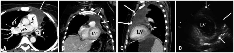
Fig. 1 Contrast-enhanced computed tomography showing a large heterogeneous anterior mediastinal mass (arrows) invading the mediastinal structures and left anterior chest wall with encirclement and compression of the main and left pulmonary arteries (A). The mass invaded the pericardium and was inseparable from the ventricular walls (B and C). Transthoracic echocardiogram parasternal short axis view showing a large mass encircling the anterior and lateral left ventricular walls (arrows) (D). LPA: left pulmonary artery, MPA: main pulmonary artery, RPA: right pulmonary artery, LV: left ventricle.
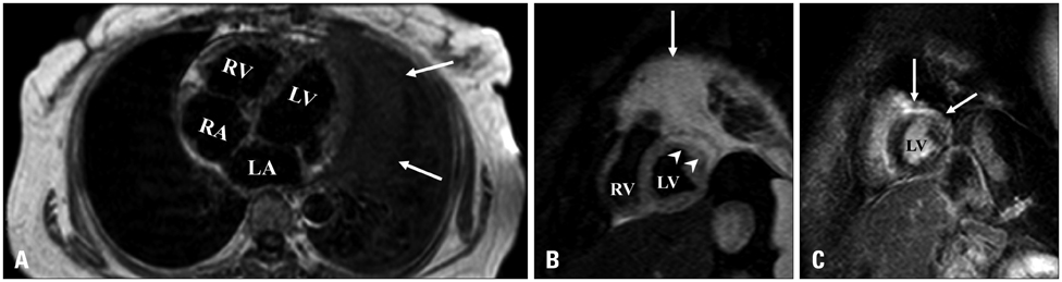
Fig. 2 A: Axial T1-weighted imaging showing the mass inseparable from the left ventricular wall (arrows). B: Short-axis T2-weighted imaging showing myocardial edema in the anterior and lateral walls of the left ventricle (arrowheads) with high signal intensity of the mass (arrow). C: Short-axis late gadolinium enhancement imaging showing non-ischemic sub-epicardial to mid wall late enhancement involving the anterior wall with extension to a small portion of anterior septum/anterolateral walls (arrows). LA: left atrium, LV: left ventricle, RA: right atrium, RV: right ventricle.
Reference
-
1. Savage KJ. Primary mediastinal large B-cell lymphoma. Oncologist. 2006; 11:488–495.
Article2. O'Mahony D, Peikarz RL, Bandettini WP, Arai AE, Wilson WH, Bates SE. Cardiac involvement with lymphoma: a review of the literature. Clin Lymphoma Myeloma. 2008; 8:249–252.3. Yang CC, Tsai HW, Lai ST, Wu HC, Lo CY, Chang Y. Mediastinal diffuse large B-cell lymphoma invading the left atrium mimicking coronary artery disease with a mural thrombus. J Chin Med Assoc. 2012; 75:606–609.
Article4. Goldman M, Matthews R, Meng H, Bilfinger T, Kort S. Evaluation of cardiac involvement with mediastinal lymphoma: the role of innovative integrated cardiovascular imaging. Echocardiography. 2012; 29:E189–E192.
Article5. Bley TA, Zeiser R, Ghanem NA, Hackanson B, Brink I, Langer M. High grade cardiac lymphoma vitality monitoring by gadolinium-enhanced magnetic resonance imaging (MRI). In Vivo. 2005; 19:689–693.
- Full Text Links
-
- Actions
-
Cited
- CITED
-
- Close
- Share
-
- Similar articles
-
- A Case of Secondary Cutaneous Diffuse Large B-cell Lymphoma
- A Case of Primary Cutaneous Diffuse Large B-cell Lymphoma
- Relapse of Ocular Lymphoma following Primary Testicular Diffuse Large B-cell Lymphoma
- Diffuse Large B-cell Lymphoma of the Sacral Nerve Root ; Presenting as a Polyradiculoneuropathy
- A case of Ki-1 positive large-cell lymphoma
