Korean J Obstet Gynecol.
2010 Dec;53(12):1129-1135. 10.5468/kjog.2010.53.12.1129.
Sclerosing stromal tumors of the ovary occurred in various ages
- Affiliations
-
- 1Department of Obstetrics and Gynecology, Soonchunhyang University Seoul Hospital, Soonchunhyang University College of Medicine, Seoul, Korea. shcha@schmc.ac.kr
- 2Department of Pathology, Soonchunhyang University Seoul Hospital, Soonchunhyang University College of Medicine, Seoul, Korea.
- KMID: 2037515
- DOI: http://doi.org/10.5468/kjog.2010.53.12.1129
Abstract
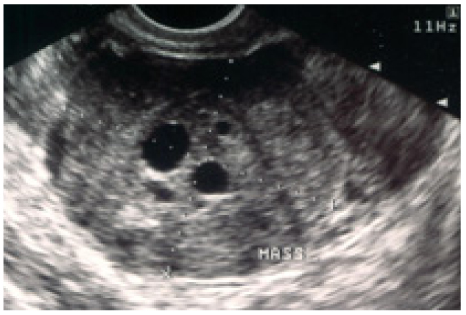
- Sclerosing stromal tumor (SST) of the ovary is a rare, benign tumor. The most common clinical symptom is menstrual irregularity. Diagnosis of SST is often made by postoperative pathologic examination. The important differential diagnoses are other sex cord stromal tumors including fibroma, thecoma and etc. We present four cases of SST of the ovary during 10 years with a brief review of the literature.
Figure
Reference
-
1. Youm HS, Cha DS, Han KH, Park EY, Hyon NN, Chong Y. A case of huge sclerosing stromal tumor of the ovary weighing 10 kg in a 71-year-old postmenopausal woman. J Gynecol Oncol. 2008. 19:270–274.2. Chalvardjian A, Scully RE. Sclerosing stromal tumors of the ovary. Cancer. 1973. 31:664–670.3. Saitoh A, Tsutsumi Y, Osamura RY, Watanabe K. Sclerosing stromal tumor of the ovary. Immunohistochemical and electron-microscopic demonstration of smooth-muscle differentiation. Arch Pathol Lab Med. 1989. 113:372–376.4. Kuscu E, Oktem M, Karahan H, Bilezikci B, Demirhan B. Sclerosing stromal tumor of the ovary: a case report. Eur J Gynaecol Oncol. 2003. 24:442–444.5. Prat J. Pathology of the ovary. 2004. Philadelphia: Saunders.6. Korczyinski J, Gottwald L, Pasz-Walczak G, Kubiak R, Bienkiewicz A. [Sclerosing stromal tumor of the ovary in a 30-year-old woman. A case report and review of the literature]. Ginekol Pol. 2005. 76:471–475.7. Ihara N, Togashi K, Todo G, Nakai A, Kojima N, Ishigaki T, et al. Sclerosing stromal tumor of the ovary: MRI. J Comput Assist Tomogr. 1999. 23:555–557.8. Lee MS, Cho HC, Lee YH, Hong SR. Ovarian sclerosing stromal tumors: gray scale and color Doppler sonographic findings. J Ultrasound Med. 2001. 20:413–417.9. Yoon GS, Kim MS. A case of sclerosing stromal tumor of the ovary. Korean J Obstet Gynecol. 2003. 46:2052–2055.10. Gee DC, Russell P. Sclerosing stromal tumours of the ovary. Histopathology. 1979. 3:367–376.11. Fox H, Wells M. Haines and Taylor Obstetrical and Gynecological Pathology. 2002. 5th ed. New York: Cherchill Livingstone.12. Terauchi F, Onodera T, Nagashima T, Kobayashi Y, Moritake T, Oharaseki T, et al. Sclerosing stromal tumor of the ovary with elevated CA125. J Obstet Gynaecol Res. 2005. 31:432–435.13. Lee HC, Kim BR, Lee JS, Lim BI, Kim HG, Moon HB, et al. A case of ovarian sclerosing stromal tumor with massive ascites and elevated CA 125. Korean J Obstet Gynecol. 2009. 52:120–124.
- Full Text Links
- Actions
-
Cited
- CITED
-
- Close
- Share
- Similar articles
-
- Sclerosing stromal cell tumor of the ovary in pregnancy: A case report and review of the literature
- Sclerosing stromal tumor of the ovary
- A Case Of Sclerosing Stromal Tumor Of The Ovary
- A Case of Sclerosing Stromal Tumor of the Ovary
- Sclerosing Stromal Tumor of the Ovary: MR-Pathologic Correlation in Three Cases

