Korean J Obstet Gynecol.
2011 Feb;54(2):119-122. 10.5468/KJOG.2011.54.2.119.
Pelvic organ prolapse and uterine inversion: A case report and literature review
- Affiliations
-
- 1Department of Obstetrics and Gynecology, Kwangju Christian Hospital, Gwangju, Korea. drsunsuh@hotmail.com
- 2Department of Anesthesiology and Pain Medicine, Kwangju Christian Hospital, Gwangju, Korea.
- KMID: 2037503
- DOI: http://doi.org/10.5468/KJOG.2011.54.2.119
Abstract
- Non-puerperal uterine inversion is a rare clinical problem with only 150 cases reported. It usually results from a tumor implanted on fundus of the uterus. Uterine inversion associated with pelvic organ prolapse (POP) is extremely rare with only 1 case reported. A 74-year-old multiparous menopausal women was admitted to our hospital with the following condition: purulent vaginal discharge, voiding difficulty, lower abdominal discomfort, vaginal bleeding, protruding vaginal mass and POP. She was diagnosed as a POP with uterine inversion. After a posterior hysterotomy, vaginal hysterectomy, bilateral salpingoophorectomy and colpocleisis was performed and the pathological examination revealed a pyometra. We report a rare case of uterine inversion associated with POP and pyometra with a brief review of literature.
MeSH Terms
Figure
-
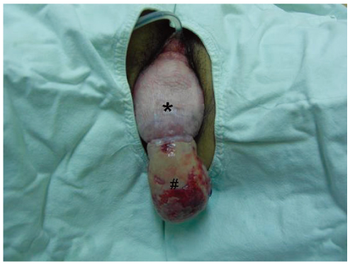
Fig. 1 View obtained at physical examination; The prolapsed structure, 18 cm in length, consisted of the vagina (*) and the inverted endometrium (#).
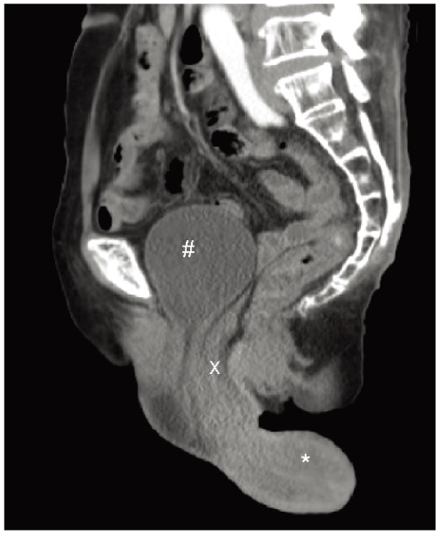
Fig. 2 Sagittal image of pelvic cavity on abdominopelvic computed tomography; a U-shaped uterine cavity (*), distended bladder (#) and small bowel herniation (x).

Fig. 3 View obtained after posterior hysterotomy; both tubes (*), round (x) and ovarian ligament (#).
Reference
-
1. Richter HE, Varner RE. Berek JS, Novak E, editors. Pelvic organ prolapse. Berek and Novak's gynecology. 2007. 14th ed. Philadelphia (PA): Lippincott Williams and Wilkins;897–929.2. Das P. Inversion of the uterus. J Obstet Gynaecol Br Emp. 1940. 47:525–548.3. Gomez-Lobo V, Burch W, Khanna PC. Nonpuerperal uterine inversion associated with an immature teratoma of the uterus in an adolescent. Obstet Gynecol. 2007. 110:491–493.4. Lupovitch A, England ER, Chen R. Non-puerperal uterine inversion in association with uterine sarcoma: case report in a 26-year-old and review of the literature. Gynecol Oncol. 2005. 97:938–941.5. Song MJ, You SH, Suh MJ, Kook IY, Yoon JH. A case of uterine inversion resulted from prolapse of huge pedunculated uterine submucosal leiomyoma. Korean J Obstet Gynecol. 2007. 50:380–383.6. Reisenauer C, Solomayer E. Images in clinical medicine. Pelvic-organ prolapse and uterine inversion. N Engl J Med. 2009. 360:1238.7. Wiedswang G, Moen MH. Non-puerperal inversion of the uterus. Acta Obstet Gynecol Scand. 1989. 68:559–560.8. Palluel V, Dargent D. Uterine inversion and prolapse. Bull Fed Soc Gynecol Obstet Lang Fr. 1966. 18:482–483.9. Pride GL, Shaffer RL. Nonpuerperal uterine inversion. Report of an unusual case. Obstet Gynecol. 1977. 49:361–364.10. Takano K, Ichikawa Y, Tsunoda H, Nishida M. Uterine inversion caused by uterine sarcoma: a case report. Jpn J Clin Oncol. 2001. 31:39–42.11. Lascarides E, Cohen M. Surgical management of nonpuerperal inversion of the uterus. Obstet Gynecol. 1968. 32:376–381.12. Hsieh TT, Lee JD. Sonographic findings in acute puerperal uterine inversion. J Clin Ultrasound. 1991. 19:306–309.13. Lewin JS, Bryan PJ. MR imaging of uterine inversion. J Comput Assist Tomogr. 1989. 13:357–359.14. Buyukkurt S, Vardar MA, Zeren H, Ozgunen FT. Non-puerperal inversion of the uterus caused by leiomyosarcoma: a case report and clinical management. J Obstet Gynaecol Res. 2007. 33:402–406.15. FitzGerald MP, Brubaker L. Colpocleisis and urinary incontinence. Am J Obstet Gynecol. 2003. 189:1241–1244.
