Korean Circ J.
2011 Apr;41(4):217-219. 10.4070/kcj.2011.41.4.217.
An Atypical Mitral Valve Prolapse in a Patient With Behcet's Disease
- Affiliations
-
- 1Cardiology Division, NHIC Ilsan Hospital, Goyang, Korea.
- 2Cardiology Division, Department of Internal Medicine, Yonsei University College of Medicine, Seoul, Korea. sejoong@yuhs.ac
- 3Cardiology Division, Department of Internal Medicine, Hallym University College of Medicine, Seoul, Korea.
- KMID: 2028948
- DOI: http://doi.org/10.4070/kcj.2011.41.4.217
Abstract
- We report the case of a 42-year-old male who was admitted to the hospital with progressive dyspnea. Cardiomegaly and diffuse pulmonary edema were visible on chest X-ray and multiple oral and genital ulcers on physical examination. On admission, echocardiography revealed mitral valve prolapse (MVP) predominantly involving a basal portion of the posterior leaflet, with severe mitral regurgitation. A successful mitral valve replacement with St. Jude #29 was performed, after pre-treatment with prednisolone for 2 weeks. Fifteen months following the operation, the patient expired from severe pulmonary edema and secondary pneumonia. This case demonstrates, for the first time in the literature, an unusual feature of mitral prolapse in the basal portion with severe mitral regurgitation in a patient with Behcet's disease. As suggested by this case, we should consider an atypical type of MVP as a possible inflammatory involvement of the heart in patients with Behcet's disease.
MeSH Terms
Figure
-
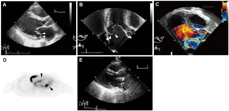
Fig. 1 A: parasternal long view showing mitral valve prolapse, predominantly involving a basal portion of the posterior mitral leaflet (white arrowheads) and aneurysmal changes of the aortic valve in TTE. B: TEE (156 degree) showing mitral valve prolapse in basal portion of posterior mitral leaflet (white arrowheads). C: TEE (136 degree) showing severe mitral regurgitation. D: PET (18-FDG) showing increased uptake along the right atrial wall and mitral annulus (black arrowheads). E: parasternal long axis view showing almost totally detached prosthetic mitral valve and displacement into the left atrium (white arrowheads).
Reference
-
1. Verma S, Mesan TG. Mitral-valve repair for mitral-valve prolapse. N Engl J Med. 2009. 361:2261–2269.2. Gurgun C, Ercan E, Ceyhan C, et al. Cardiovascular involvement in Behçet's disease. Jpn Heart J. 2002. 43:389–398.3. James DG. 'Silk Route disease' (Behçet's disease). West J Med. 1988. 148:433–437.4. Cheng TO. Behçet's disease ('Silk Route disease') and mitral valve prolapse. West J Med. 1989. 150:91.5. Shen LL, Cui GG, Liang RL. Valve prolapse in Behçet's disease. Br Heart J. 1985. 54:100–101.6. Park K, Song J, Kang D, et al. Clinical characteristics of patients with cardiac involvement in Behçet's disease. Korean Circ J. 2005. 35:847–853.
