Comparative Analysis of Two Cementless Stems in Total Hip Arthroplasties in Patients with Osteonecrosis of Femoral Head: Summit(R) Stem and Bencox(R) Stem
- Affiliations
-
- 1Department of Orthopaedic Surgery, College of Medicine, The Catholic University of Korea, Seoul, Korea. sykwon@catholic.ac.kr
- KMID: 2014894
- DOI: http://doi.org/10.5371/jkhs.2012.24.1.25
Abstract
- PURPOSE
We compared the clinical and radiological outcomes of total hip arthroplasty (THR) using Summit and Bencox stems.
MATERIALS AND METHODS
The patients who underwent cementless total hip arthroplasty were recruited with a satisfactory condition of a minimum three years of follow-ups after THR. Those patients were divided into two groups, those with Summit stems and those with Bencox stems. Summit stems were in 36 patients(40 hips), and Bencox stems in 36 patients(48 hips). Summit and Bencox stems had 78 months and 42.2 months as a mean follow-up, respectively. The clinical and radiological evaluations of femoral components were performed.
RESULTS
There was no difference in clinical results between the two groups. Under the radiological findings, there were no osteolytic changes or loosening. Osseointegration was detected at an average of 6.4 months(3-12 months) in the Bencox stem on the distal portion of the femoral stem, and cortical hypertrophy was detected on 6 hips with a Summit stem.
CONCLUSION
The clinical and radiological evaluations in both systems showed excellent outcomes at the three year follow-ups, and there was no statistical difference on the clinical and radiological results between the two groups. Thigh pain and cortical hypertrophy were not detected in the Bencox stem, and that wound would be caused by surface treatment methods of the femoral stem, and morphological differences.
Figure
-
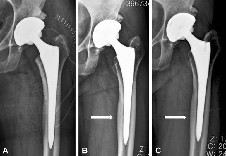
Fig. 1 A 55-years-old woman. (A) A postoperative AP radiograph, (B) AP radiograph taken at the 12 months followup begins to show cortical hypertrophy at the distal portion of stem (white arrow), and the patient complained of thigh pain. (C) AP radiograph taken at the 36 months followup shows the cortical hypertrophy precisely (white arrow).
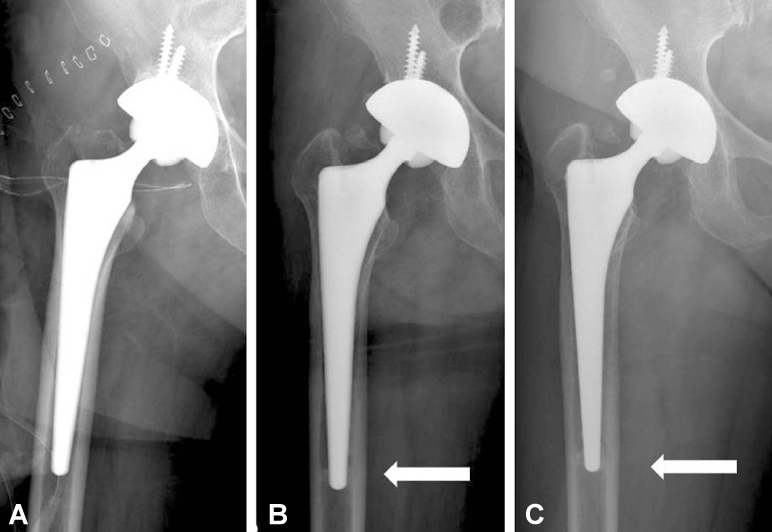
Fig. 2 A 63-years-old man. (A) A postoperative AP radiograph, (B) AP radiograph taken at the 6 months followup begins to show prosthesis to bone union at the distal portion of stem (white arrow). (C) AP radiograph taken at the 36 months followup shows prosthesis to bone union precisely (white arrow).
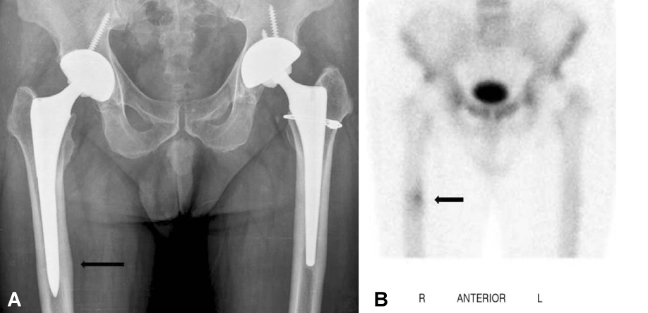
Fig. 3 A 58-years-old man. His diagnosis is osteonecrosis of femoral head. On the right side, he was performed THR using Summit stem 6 years ago, and on the left side, performed THR using BENCOX stem 3 years ago. He complained of right thigh pain. (A) A postoperative AP radiograph shows right proximal femur cortical hypertrophy (arrow). (B) A whole body bone scan shows hot uptake on the right proximal femur (arrow).
Cited by 3 articles
-
Minimum Seven-year Follow-up of Cementless Total Hip Arthroplasty with the COREN Hip System
Sang-Min Kim, Young-Wan Moon, Seung-Jae Lim, Jae-Won Heo, Yong-Sik Kim, Young-Wook Lim, Youn-Soo Park
Hip Pelvis. 2013;25(3):173-181. doi: 10.5371/hp.2013.25.3.173.Primary Total Hip Arthroplasty Using Summit® Stems in Korean: Minimum Four-year Follow-up
Jae Sik Yoon, Joon Sun Kang, Kyoung Ho Moon
Hip Pelvis. 2017;29(4):228-233. doi: 10.5371/hp.2017.29.4.228.Cementless Total Hip Arthroplasty Using the COREN Hip System: A Minimum Five-Year Follow-up Study
Hee Joong Kim, Jeong Joon Yoo, Wonyeong Seo, Min Nyeon Kim, Taehoon Kang
Hip Pelvis. 2018;30(3):162-167. doi: 10.5371/hp.2018.30.3.162.
Reference
-
1. Brinker MR, Rosenberg AG, Kull L, Galante JO. Primary total hip arthroplasty using noncemented porous-coated femoral components in patients with osteonecrosis of the femoral head. J Arthroplasty. 1994. 9:457–468.
Article2. Buser D, Schenk RK, Steinemann S, Fiorellini JP, Fox CH, Stich H. Influence of surface characteristics on bone integration of titanium implants. A histomorphometric study in miniature pigs. J Biomed Mater Res. 1991. 25:889–902.
Article3. Anselme K. Osteoblast adhesion on biomaterials. Biomaterials. 2000. 21:667–681.
Article4. Lim YW, Kwon SY, Sun DH, Kim HE, Kim YS. Enhanced cell integration to titanium alloy by surface treatment with microarc oxidation: a pilot study. Clin Orthop Relat Res. 2009. 467:2251–2258.
Article5. Zhu X, Kim KH, Jeong Y. Anodic oxide films containing Ca and P of titanium biomaterial. Biomaterials. 2001. 22:2199–2206.
Article6. Callaghan JJ, Dysart SH, Savory CG. The uncemented porous-coated anatomic total hip prosthesis. Two-year results of a prospective consecutive series. J Bone Joint Surg Am. 1988. 70:337–346.
Article7. Kim YH, Kim VE. Cementless porous-coated anatomic medullary locking total hip prostheses. J Arthroplasty. 1994. 9:243–252.
Article8. Annaz B, Hing KA, Kayser M, Buckland T, Di Silvio L. Porosity variation in hydroxyapatite and osteoblast morphology: a scanning electron microscopy study. J Microsc. 2004. 215:100–110.
Article9. Li LH, Kong YM, Kim HW, et al. Improved biological performance of Ti implants due to surface modification by micro-arc oxidation. Biomaterials. 2004. 25:2867–2875.
Article10. Kim YS, Kwon SY, Sun DH, Han SK, Maloney WJ. Modified posterior approach to total hip arthroplasty to enhance joint stability. Clin Orthop Relat Res. 2008. 466:294–299.
Article11. Harris WH. Traumatic arthritis of the hip after dislocation and acetabular fractures: treatment by mold arthroplasty. An end-result study using a new method of result evaluation. J Bone Joint Surg Am. 1969. 51:737–755.
Article12. Khalily C, Lester DK. Results of a tapered cementless femoral stem implanted in varus. J Arthroplasty. 2002. 17:463–466.
Article13. Martell JM, Pierson RH 3rd, Jacobs JJ, Rosenberg AG, Maley M, Galante JO. Primary total hip reconstruction with a titanium fiber-coated prosthesis inserted without cement. J Bone Joint Surg Am. 1993. 75:554–571.
Article14. Engh CA, Massin P, Suthers KE. Roentgenographic assessment of the biologic fixation of porous-surfaced femoral components. Clin Orthop Relat Res. 1990. 257:107–128.
Article15. Engh CA, Hooten JP Jr, Zettl-Schaffer KF, et al. Porous-coated total hip replacement. Clin Orthop Relat Res. 1994. 298:89–96.
Article16. Gruen TA, McNeice GM, Amstutz HC. "Modes of failure" of cemented stem-type femoral components: a radiographic analysis of loosening. Clin Orthop Relat Res. 1979. 141:17–27.17. Brooker AF, Bowerman JW, Robinson RA, Riley LH Jr. Ectopic ossification following total hip replacement. Incidence and a method of classification. J Bone Joint Surg Am. 1973. 55:1629–1632.18. Lee TM, Tsai RS, Chang E, Yang CY, Yang MR. The cell attachment and morphology of neonatal rat calvarial osteoblasts on the surface of Ti-6Al-4V and plasma-sprayed HA coating: effect of surface roughness and serum contents. J Mater Sci Mater Med. 2002. 13:341–350.19. Yang IH, Park SH, Han CD. Cementless total hip arthroplasty using Ceramic-on-ceramic Summit® System: 3 years follow up. J Korean Hip Soc. 2007. 19:9–15.
Article20. Khanuja HS, Vakil JJ, Goddard MS, Mont MA. Cementless femoral fixation in total hip arthroplasty. J Bone Joint Surg Am. 2011. 93:500–509.
Article21. Schreiner U, Scheller G, Herbig J, Jani L. Mid-term results of the cementless CLS stem. A 7- to 11-year follow-up study. Arch Orthop Trauma Surg. 2001. 121:321–324.
- Full Text Links
-
- Actions
-
Cited
- CITED
-
- Close
- Share
-
- Similar articles
-
- Result of a Minimum Five-Year Follow-Up of Hip Arthroplasty Using the Bencox(R) Hip Stem
- Ceramic-on-Ceramic Bearing Total Hip Arthroplasty: Five-Year Midterm Follow-up Results
- Ultra-Short Bone Conserving Cementless Femoral Stem
- 10-15 Year Follow-up Results of Total Hip Arthroplasty Using a CLS (Cementless Spotorno) Stem for Osteonecrosis of the Femoral Head
- Cementless Total Hip Arthroplasty with Circumferentially Proximal Porous-Coated Femoral Stem -Minimum 10-Year Follow-up Results
