Ovarian dysgerminoma associated with pregnancy
- Affiliations
-
- 1Department of Obstetrics and Gynecology, St. Vincent's Hospital, The Catholic University of Korea School of Medicine, Suwon, Korea. dcpark@catholic.ac.kr
- 2Department of Pathology, St. Vincent's Hospital, The Catholic University of Korea School of Medicine, Suwon, Korea.
- KMID: 2013245
- DOI: http://doi.org/10.5468/KJOG.2011.54.4.218
Abstract
- A case of a 33-year-old 3 gravida, 1 para pregnant woman in the 7th week of gestation who was concurrented with dysgerminoma is presented. Measurements were made on serum lactic dehydrogenase, alpha-fetoprotein, CA-125, and pelvicmagnetic resonance imaging. The clinical stage was IA and right salpingo-oophorectomy and biopsy of the left ovary were done preserving the pregnancy. A cytological finding was non-specific. The frequency of malignant ovarian tumors associated with pregnancy and the treatment for it were discussed.
Keyword
MeSH Terms
Figure
-
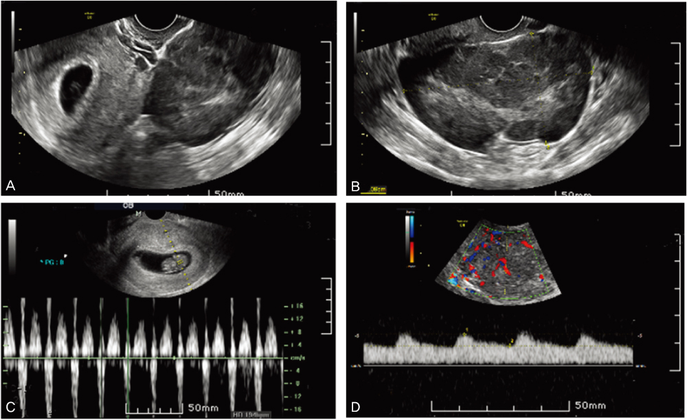
Fig. 1 Ultrasonographic findings. (A). Fetal heart rate is normal (B). The mass had multilocular profiles with some solid parts, and its wall looked slightly thick (C). The vascularization of tumor mass was checked and the resistance index (RI) was calculated. RI=0.33 (D).
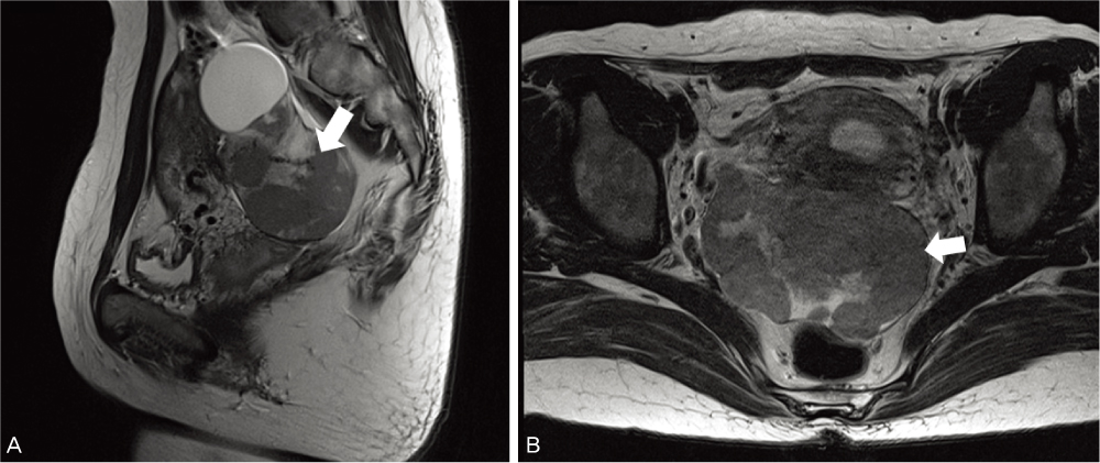
Fig. 2 Pelvic magnetic resonance imaging without enhance was perfomed. It measured a 5.8×9.8 cm in sized and was a lobulating contoured mass with intermediate signal intensity in T2 images. And a 4.3×3.3 cm sized cystic lesion was also observed. (A) Sagittal view. (B) Axial view. Arrows: dysgerminoma.

Fig. 3 Laparoscopic finding. Laparoscopic right adnexectomy and contralateral ovary biopsy were performed.

Fig. 4 Microscopically, monotonous polygonal cells form well defined nests separated by fibrous stroma (A: H&E, ×100). In certain area tumors cells form cords or trabecular patterns (B: H&E, ×400).
Reference
-
1. Creasman WT, Rutledge F, Smith JP. Carcinoma of the ovary associated with pregnancy. Obstet Gynecol. 1971. 38:111–116.2. Beischer NA, Buttery BW, Fortune DW, Macafee CA. Growth and malignancy of ovarian tumours in pregnancy. Aust N Z J Obstet Gynaecol. 1971. 11:208–220.3. Park BW, Song SS, Lee HD. A case of a huge dysgerminoma of the ovary. Korean J Obstet Gynecol. 1983. 26:109–111.4. Kim SY, Kim YS, Park YH, Jeon K, Lim SG, Kim JW. A case of recurrent ovarian dysgerminora. Korean J Obstet Gynecol. 1983. 26:965–967.5. Kim JY, Choi SL, Park IW, Jun HC, Jung DS, Jo JD, et al. A case of pure dysgerminoma with syncytiotrophoblastic giant cell secreating hCG. Korean J Obstet Gynecol. 2003. 46:469–473.6. Bakri YN, Ezzat A, Akhtar , Dohami , Zahrani . Malignant germ cell tumors of the ovary. Pregnancy considerations. Eur J Obstet Gynecol Reprod Biol. 2000. 90:87–91.7. DiSaia PJ, Townsend DE, Morrow CP. The rationale for less than radical treatment for gynecologic malignancy in early reproductive years. Obstet Gynecol Surv. 1974. 29:581–593.8. Munnell EW. Primary ovarian cancer associated with pregnancy. Clin Obstet Gynecol. 1963. 30:983–993.9. Graber EA, Barber HR. Barber HR, Graber EA, editors. Ovarian tumors in pregnancy. Surgical disease in pregnancy. 1974. Philadelphia (PA): W.B. Saunders;428–439.
- Full Text Links
-
- Actions
-
Cited
- CITED
-
- Close
- Share
-
- Similar articles
-
- A Case of Pure Dysgerminoma with Syncytiotrophoblastic Giant Cell Secreating HCG
- Torsion and ruptured dysgerminoma of ovary in pregnancy
- Radiation Therapy of Ovarian Dysgerminoma
- Pelviscopic Diagnosis and Treatment of Two Cases of Ovarian Pregnancy
- A Case of Dysgerminoma Incidentally Found after Pelviscopic Ovarian Surgery
