J Korean Soc Radiol.
2010 May;62(5):461-465. 10.3348/jksr.2010.62.5.461.
Diagnostic Usefulness of a Multimodality Study in a Mediastinal Hemangioma: A Case Report
- Affiliations
-
- 1Department of Radiology, Soonchunhyang University Hospital, Korea.
- 2Department of Radiology, Soonchunhyang Chunan Hospital, Korea. ytokim@schch.co.kr
- KMID: 2002954
- DOI: http://doi.org/10.3348/jksr.2010.62.5.461
Abstract
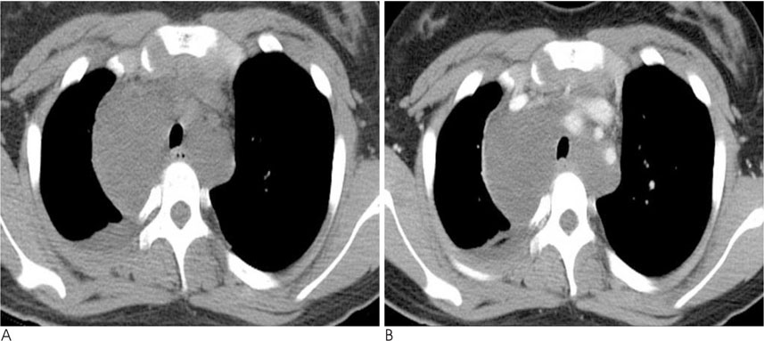
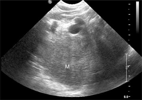
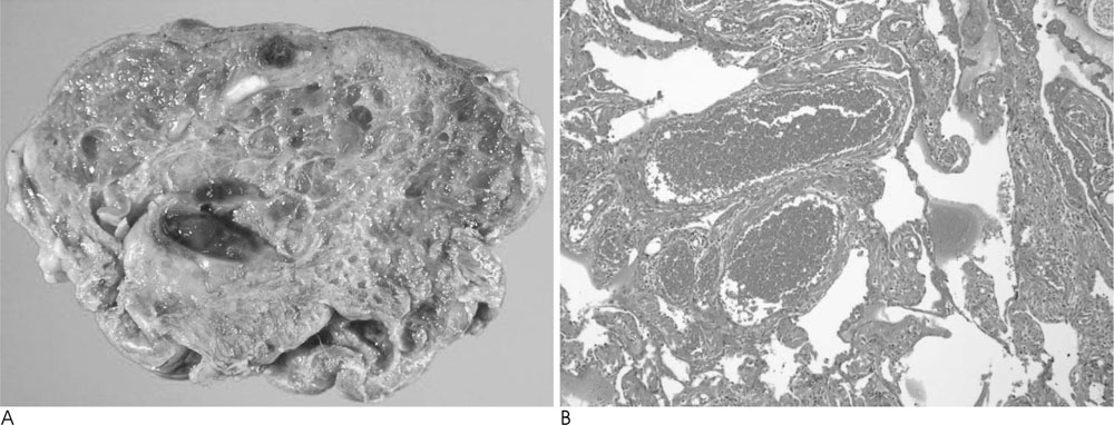
- Cavernous hemangiomas of the mediastinum are rare tumors that usually occur in young patents. We present a case of cavernous hemangioma in a 12-year-old girl. This case was seen as nonenhancing cystic mass on CT, mimicking lymphangioma. US demonstrated a hyperechoic mass, while MRI revealed a T2 high SI with delayed homogenous enhancement. US and MRI were found to be useful tools for the diagnosis of hemangiomas, seen as a cystic mass on a chest CT scan.
MeSH Terms
Figure
Reference
-
1. Cohen AJ, Sbaschnig RJ, Hochholzer L, Lough FC, Albus RA. Mediastinal hemangiomas. Ann Thorac Surg. 1987; 43:656–659.2. Davis JM, Mark GJ, Greene R. Benign blood vascular tumors of the mediastinum: report of four cases and review of the literature. Radiology. 1978; 126:581–587.3. McAdams HP, Rosado-de-Christenson ML, Moran CA. Mediastinal hemangioma: radiographic and CT features in 14 patients. Radiology. 1994; 193:399–402.4. Semisa M, Fiore D, Ravasini R, Macchi C. Computed tomography of mediastinal cavernous hemangioma: a case report. Rays. 1986; 11:31–33.5. Bree RL, Schwab RE, Glazer GM, Fink-Bennett D. The varied appearances of hepatic cavernous hemangiomas with sonography, computed tomography, magnetic resonance imaging and scintigraphy. Radiographics. 1987; 7:1153–1175.6. Agarwal PP, Seely JM, Matzinger FR. Case 130: mediastinal hemangioma. Radiology. 2008; 246:634–637.7. Fujimoto K, Muller NL. Anterior mediastinal masses. In : Muller NL, Silva CI, editors. Imaging of the chest, volume II. Philadelphia: Saunders;2008. p. 1512.8. Ishii K, Maeda K, Hashihira M, Miyamoto Y, Kanegawa K, Kusumoto M, et al. MRI of mediastinal cavernous hemangioma. Pediatr Radiol. 1990; 20:556–557.9. Abe K, Akata S, Ohkubo Y, Park J, Kakizaki D, Simatani H, et al. Venous hemangioma of the mediastinum. Eur Radiol. 2001; 11:73–75.10. Nchimi A, Ghaye B, Szapiro D, Thiry A, Dondelinger RF. A complex anterior mediastinal mass: demonstration of pericardial haemangioma by dynamic MRI (2003:10b). Eur Radiol. 2004; 14:160–163.


