J Korean Acad Prosthodont.
2013 Jul;51(3):167-174. 10.4047/jkap.2013.51.3.167.
Evaluation of titanium surface properties by Nd: YVO4 laser irradiation: pilot study
- Affiliations
-
- 1Department of Prosthodontics, School of Dentistry, Chonnam National University, Gwangju, Korea. psw320@chonnam.ac.kr
- 2Department of Dental Science, School of Dentistry, Chonnam National University, Gwangju, Korea.
- KMID: 2000165
- DOI: http://doi.org/10.4047/jkap.2013.51.3.167
Abstract
- PURPOSE
This study was conducted to evaluate the roughness and surface alternations of three differently blasted titanium discs treated by Nd: YVO4 Laser irradiation in different conditions.
MATERIALS AND METHODS
Thirty commercially pure titanium discs were prepared and divided into three groups. Each group was consisted of 10 samples and blasted by ZrO2 (zirconium dioxide), Al2O3 (aluminum oxide), and RBM (resorbable blasted media). All the samples were degreased by ultrasonic cleaner afterward. Nine different conditions were established by changing scanning speed (100, 300, 500 mm/s) and repetition rate (5, 15, 35 kHz) of Nd: YVO4 Laser (Laser Pro D-20, Laserval Korea(R) Seoul, South Korea). After laser irradiation, a scanning electron microscope, X-ray diffraction analysis, energy dispersive X-ray spectroscopic analysis, and surface roughness analysis were used to assess the roughness and surface alternations of the samples.
RESULTS
According to a scanning electron microscope (SEM), titanium discs treated with laser irradiation showed characteristic patterns in contrast to the control which showed irregular patterns. According to the X-ray diffraction analysis, only Al2O3 group showed its own peak. The oxidation tendency and surface roughness of titanium were similar to the control in the energy dispersive X-ray spectroscopic analysis. The surface roughness was inversely proportional to the scanning speed, whereas proportional to the repetition rate of Nd: YVO4.
CONCLUSION
The surface microstructures and roughness of the test discs were modified by the radiation of Nd: YVO4 laser. Therefore, laser irradiation could be considered one of the methods to modify implant surfaces for the enhancement of osseointegration.
Keyword
MeSH Terms
Figure
-
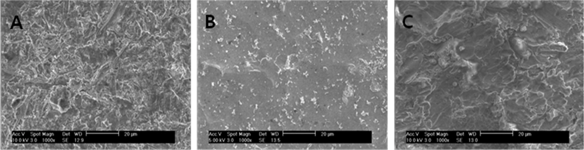
Fig. 1. SEM micrographs of titanium disc without laser irradiation at original magnification of ×1,000. A: group Z, B: group A, C: group R.
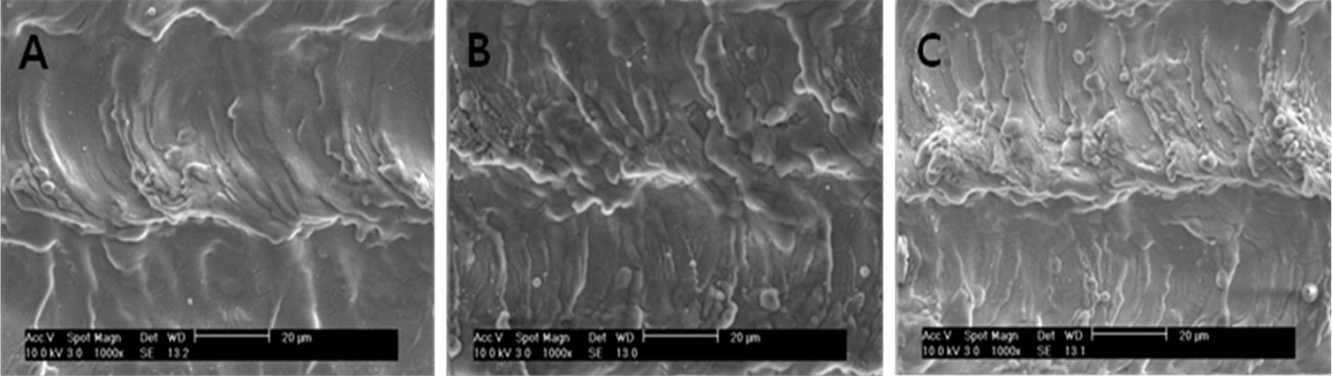
Fig. 2. SEM micrographs of titanium disc by laser irradiation at original magnification of ×1,000. A: bead form in group Z, B: wave-like pattern in group A, C: parallel scratch in group R. ∗ Specimen number is S1R35.
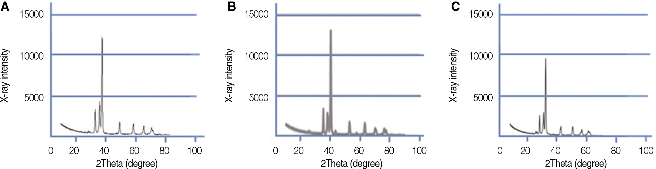
Fig. 3. The XRD spectra of the Ti specimen surfaces. A: group Z, B: group A, C: group R.
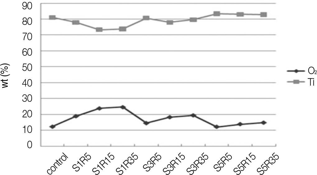
Fig. 4. The result of EDX analysis in group Z.
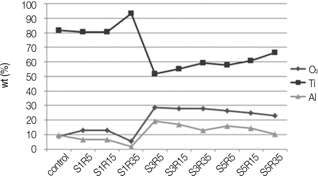
Fig. 5. The result of EDX analysis in group A.
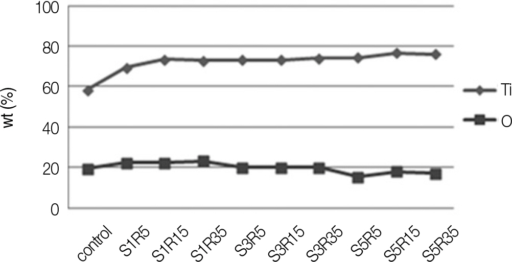
Fig. 6. The result of EDX analysis in group R.
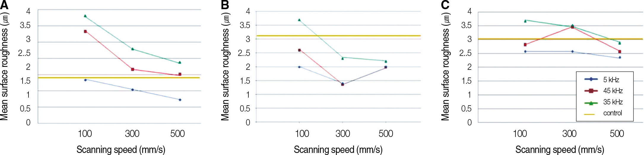
Fig. 7. Changes of surface roughness by Scanning Speed and Repetition rate: A, group Z; B, group A; C, group R.
Reference
-
1.Albrektsson T., Lekholm U. Osseointegration: current state of the art. Dent Clin North Am. 1989. 33:537–54.2.Wennerberg A., Albrektsson T. Effects of titanium surface topography on bone integration: a systematic review. Clin Oral Implants Res. 2009. 20:172–84.
Article3.Mendonça G., Mendonça DB., Aragão FJ., Cooper LF. Advancing dental implant surface technology-from micron- to nanotopog-raphy. Biomaterials. 2008. 29:3822–35.4.Marchi J., Delfino CS., Bressiani JC., Bressiani AHA., Marques MM. Cell proliferation of human fibroblasts on alumina and hydroxyapatite-based ceramics with different surface treatments. Int J Appl Ceram Technol. 2010. 7:139–47.
Article5.Le Gue′hennec L., Soueidan A., Layrolle P., Amouriq Y. Surface treatments of titanium dental implants for rapid osseointegration. Dent Mater. 2007. 23:844–54.6.Elias CN., Oshida Y., Lima JH., Muller CA. Relationship between surface properties (roughness, wettability and morphology) of titanium and dental implant removal torque. J Mech Behav Biomed Mater. 2008. 1:234–42.
Article7.Wennerberg A., Albrektsson T., Andersson B., Krol JJ. A histomorphometric and removal torque study of screw-shaped titanium implants with three different surface topographies. Clin Oral Implants Res. 1995. 6:24–30.8.Vasanthan A., Kim H., Drukteinis S., Lacefield W. Implant surface modification using laser guided coatings: in vitro comparison of mechanical properties. J Prosthodont. 2008. 17:357–64.
Article9.Romanos GE., Everts H., Nentwig GH. Effects of diode and Nd: YAG laser irradiation on titanium discs: a scanning electron microscope examination. J Periodontol. 2000. 71:810–5.10.Stubinger S., Etter C., Miskiewicz M., Homann F., Saldamli B., Wieland M., Sader R. Surface alterations of polished and sandblasted and acid-etched titanium implants after Er: YAG, carbon dioxide, and diode laser irradiation. Int J Oral Maxillofac Implants. 2010. 25:104–11.11.Bereznai M., Pelso¨czi I., To′th Z., Turzo′ K., Radnai M., Bor Z., Fazekas A. Surface modifications induced by ns and sub-ps excimer laser pulses on titanium implant material. Biomaterials. 2003. 24:4197–203.
Article12.Heinrich A., Dengler K., Koerner T., Haczek C., Deppe H., Stritzker B. Laser-modified titanium implants for improved cell adhesion. Lasers Med Sci. 2008. 23:55–8.
Article13.Braga Francisco JC., Marques Rodrigo FC., Filho Edson de A., Guastaldi Antonio C. Surface modification of Ti dental implants by Nd: YVO4 laser irradiation. Appl Surf Sci. 2007. 253:9203–8.14.Taira T., Mukai A., Nozawa Y., Kobayashi T. Single-mode oscillation of laser-diode-pumped Nd: YVO4 microchip lasers. Opt Lett. 1991. 16:1955–7.15.Block CM., Mayo JA., Evans GH. Effects of the Nd: YAG dental laser on plasma-sprayed and hydroxyapatite-coated titanium dental implants: surface alteration and attempted sterilization. Int J Oral Maxillofac Implants. 1992. 7:441–9.16.Fields RA., Birnbaum M., Fincher CL. Highly efficient Nd: YVO4 diode-laser end-pumped laser. Appl Phys Lett. 1987. 51:1885.17.Johansson CB., Albrektsson T. A removal torque and histomorphometric study of commercially pure niobium and titanium implants in rabbit bone. Clin Oral Implants Res. 1991. 2:24–9.
Article18.Gottlander M., Albrektsson T. Histomorphometric analyses of hydroxyapatite-coated and uncoated titanium implants. The importance of the implant design. Clin Oral Implants Res. 1992. 3:71–6.
Article19.Cooper LF. A role for surface topography in creating and maintaining bone at titanium endosseous implants. J Prosthet Dent. 2000. 84:522–34.
Article20.R⊘nold HJ., Lyngstadaas SP., Ellingsen JE. Analysing the optimal value for titanium implant roughness in bone attachment using a tensile test. Biomaterials. 2003. 24:4559–64.
Article21.Kreisler M., Go¨tz H., Duschner H. Effect of Nd: YAG, Ho: YAG, Er: YAG, CO2, and GaAIAs laser irradiation on surface properties of endosseous dental implants. Int J Oral Maxillofac Implants. 2002. 17:202–11.22.Chu RT., Watanabe L., White JM., Marshal GW., Marshal SJ., Hutton JE. Temperature rise and surface modification of lased titanium cylinders. J Dent Res. 1992. 71:144. (Abstract 312).23.Peto¨ G., Karacs A., Pa′szti Z., Guczi L., Divinyi T., Joo′b A. Surface treatment of screw shaped titanium dental implants by high intensity laser pulses. Appl Surf Sci. 2002. 186:7–13.
- Full Text Links
-
- Actions
-
Cited
- CITED
-
- Close
- Share
-
- Similar articles
-
- Surface morphology characterization of laser-induced titanium implants: lesson to enhance osseointegration process
- Scanning electron microscopic study of implant surface after Er,Cr:YSGG laser irradiation
- TEMPERATURE CHANGES OF IMPLANT SURFACE IN SECOND STAGE SURGERY WITH DETAL LASER : IN VITRO STUDY
- Effect of erbium-doped yttrium-aluminum-garnet laser on surface characteristics and biocompatibility of resorbable blast media titanium
- Wettability and cellular response of UV light irradiated anodized titanium surface
