Ancient Schwannoma of the Thigh mimicking a Plexiform Malignant Peripheral Nerve Sheath Tumor: A Case Report
- Affiliations
-
- 1Department of Radiology, Daejeon St. Mary's Hospital, The Catholic University of Korea. yslee1074@medimail.co.kr
- 2Department of Orthopedic Surgery, Daejeon St. Mary's Hospital, The Catholic University of Korea.
- 3Department of Pathology, Daejeon St. Mary's Hospital, The Catholic University of Korea.
- KMID: 2000067
- DOI: http://doi.org/10.13104/jksmrm.2011.15.2.170
Abstract
- Ancient schwannoma is a rare variant of schwannoma and a slow growing benign tumor associated with degeneration that may be diagnosed as a malignant tumor, because it presents with a large size and an inhomogeneous signal intensity. The main differential diagnosis of plexiform soft tissue tumor includes plexiform neurofibroma, malignant peripheral nerve sheath tumor (MPNST). In this case, we describe the MRI findings in a case of ancient schwannoma involving left thigh of a 63-year-old woman mimicking a plexiform MPNST. The tumor appeared as an inhomogeneous signal intensity and multinodular appearance, causing misdiagnosis as a plexiform MPNST.
Keyword
MeSH Terms
Figure
-
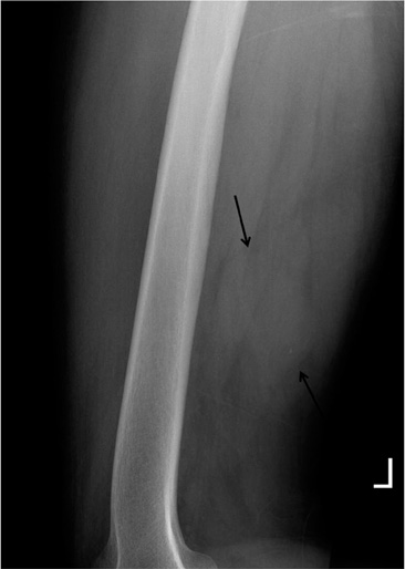
Fig. 1 Plain lateral radiograph of the left femur shows a noncalcified soft tissue mass (arrows) in the posterior thigh.
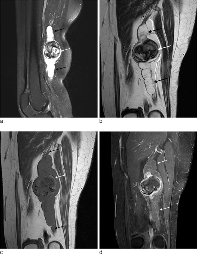
Fig. 2 Sagittal fat suppressed T2-weighted (a) and coronal T2-weighted (b) MR images show heterogeneous signal intensity mass (white arrow) associated with the multinodular, non fat-suppressed high signal lesions with proximal and distal extension in the sciatic nerve (arrows). The mass shows heterogeneous low signal intensity with mixed high signal intensity and peripheral dark signal rim (white arrow) on coronal T2 and T1-weighted image (b, c). Longitudinal extension of the low signal intensity fusiform or multinodular lesions adjacent to the mass along the sciatic nerve (arrows) on coronal T1-weighted image. Contrast enhanced coronal fat suppressed T1-weighted image (d) shows heterogeneous nodular and peripheral enhancement of the mass including non enhanced focus. However, plexiform-multinodular lesions along the sciatic nerve show no enhancement (white arrows).
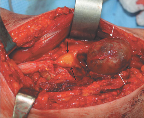
Fig. 3 Intraoperative photograph demonstrates an encapsulated mass (white arrows) associated with proximal extending multinodular yellowish lesion (black arrows) involving the sciatic nerve.
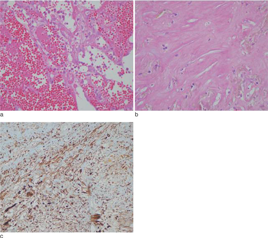
Fig. 4 Photomicrograph of the encapsulated mass demonstrates hemorrhage (a), fibrosis (b), myxoid change and inflammatory cell infiltrate (hematoxylin and eosin staining, ×100). This tumor is positive immunostaining for S100 protein (c).
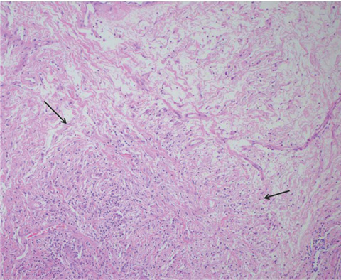
Fig. 5 Photomicrograph of multinodular yellowish lesion shows myxoid change and inflammatory cell infiltrate (arrows) in the nerve tissue (hematoxylin and eosin staining, ×100).
Reference
-
1. Woertler K. Tumors and tumor-like lesions of peripheral nerves. Semin Musculoskelet Radiol. 2010. 14:547–558.2. Isobe K, Shimizu T, Akahane T, Kato H. Imaging of ancient schwannoma. AJR Am J Roentgenol. 2004. 183:331–336.3. Lee YS, Kim JO, Park SE. Ancient schwannoma of the thigh mimicking a malignant tumour: a report of two cases, with emphasis on MRI findings. Br J Radiol. 2010. 83:e154–e157.4. Agaram NP, Prakash S, Antonescu CR. Deep-seated plexiform schwannoma: a pathologic study of 16 cases and comparative analysis with the superficial variety. Am J Surg Pathol. 2005. 29:1042–1048.5. Koo HY, Lee MJ, Lee CJ, Yoo JH. Neurilemmoma at Rare Sites: Two Cases Report. J Korean Radiol Soc. 2001. 45:571–574.6. Kransdorf MJ, Murphey MD. Neurogenic tumors. Imaging of soft tissue tumors. 2006. 2nd ed. Philadelphia: Lippncott Williams & Wilkins;328–380.7. Enzinger FM, Weiss SW. Benign tumors of peripheral nerves. Soft tissue tumors. 1995. 3rd ed. St Louis: Mosby;821–888.8. Dahl I. Ancient neurilemoma (schwannoma). Acta Pathol Microbiol Scand Sect A. 1977. 85:812–818.9. Klijanienko J, Caillaud JM, Lagace R. Cytohistologic correlations in schwannomas (neurilemmomas), including "ancient", cellular, and epithelioid variants. Diagn Cytopathol. 2006. 34:517–522.10. Hébert-Blouin MN, Amrami KK, Scheithauer BW, Spinner RJ. Multinodular/plexiform (multifascicular) schwannomas of major peripheral nerves: an underrecognized part of the spectrum of schwannomas. J Neurosurg. 2010. 112:372–382.
- Full Text Links
-
- Actions
-
Cited
- CITED
-
- Close
- Share
-
- Similar articles
-
- Two Cases of Plexiform Schwannoma
- Huge Plexiform Schwannoma of the Ulnar Nerve
- A Case of Ancient Schwannoma Masquerading as Originating from Cervical Spinal Nerve
- A Case of Ancient Schwannoma of the Submandibular Gland
- Multiple Plexiform Schwannomas Associated with Neurofibromatosis Type 2: A case report
