A Comparison Study on Human Brain Volume of White Matter, Gray Matter and Hippocampus Depending on Magnetic Resonance Imaging Conditions and Applied Brain Template
- Affiliations
-
- 1Department of Biomedical Engineering and FIRST/UHRC, Inje University, Korea. mcw@inje.ac.kr
- 2Department of Diagnostic Radiology, Haeundae Paik Hospital, Korea.
- 3Department of Psychiatry, Medical School, Inje University, Haeundae Paik Hospital, Korea.
- 4Department of Diagnostic Radiology, Medical School, Inje University, Haeundae Paik Hospital, Korea.
- KMID: 2000053
- DOI: http://doi.org/10.13104/jksmrm.2011.15.3.242
Abstract
- PURPOSE
The aim of this study was to examine the volume differences of human brain 3-D MR images obtained by automatic segmentation methods depending on brain templates and image acquisition conditions, respectively.
MATERIALS AND METHODS
3D T1-weighted MR images oriented in coronal and sagittal plane were acquired from eight healthy subjects (29.5+/-5.66 years) using two identical 3T MR scanners at different sites. Caucasian brain template and Korean elderly brain template were applied for the same subject to segment brain structural region. Volumetric differences and variation of gray matter, white matter and hippocampus depending on scan orientations and brain template types were statistically evaluated.
RESULTS
Volumetric measurements have some different results for the same subject images depending on scan orientation in identical MR scanners but not significantly. However, all segmented volumes relied upon brain templates were significantly different (p<0.05). Small variation of the volume in gray matter, white matter (coefficient of variation, CV< or =1%) and hippocampus (CV< or =3%) were obtained. Comparing the mean CV in all segmented regions, variation of scan orientation was not significantly different with inter scanner variation but variation relied upon brain templates were significantly different (p<0.001).
CONCLUSION
Authors found that brain template regarding the specific properties of the subjects is required to reduce the errors of brain volumetry.
Keyword
MeSH Terms
Figure
-

Fig. 1 Segmentation and volumetry procedure of hippocampus. (a) T1-weighted 3D MR image of human brain. (b) Segmented GM region, (c) Segmented hippocampus region and (d) its 3D ROI rendering.

Fig. 2 Differences in the volumes of (a) brain tissues and (b) hippocampus segmented from automatic processes using MNI 152 templates (circle) and Korean elderly brain templates (triangle). Results of each subject are represented as mean volumes on the image acquisition conditions *Significant difference (p<0.05) between segmented volumes **Significant difference (p<0.01) between segmented volumes

Fig. 3 (a) Scan orientation variations and (b) inter-scanner variations on measured volume of human brain tissues(GM, WM, left and right hippocampus) for each subject using MNI 152 brain template and FIRST models for hippocampus
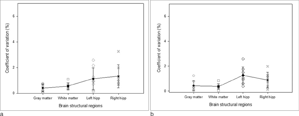
Fig. 4 (a) Scan orientation variations and (b) inter-scanner variations on measured volume of human brain tissues(GM, WM, left and right hippocampus) for each subject using Korean elderly brain/hippocampus template

Fig. 5 Segmented gray matter images overlaid with (a~c) hippocampus regions derived from FSL using FIRST models for hippocampus and (d~f) hippocampus regions derived from SPM using Korean elderly hippocampus template
Reference
-
1. Iwasaki N, Hamano K, Okada Y, et al. Volumetric quantification of brain development using MRI. Neuroradiology. 1997. 39:841–846.2. Vita A, De Peri L, Silenzi C, Dieci M. Brain morphology in first-episode schizophrenia: a meta-analysis of quantitative magnetic resonance imaging studies. Schizophrenia Research. 2006. 82:75–88.3. Utter AA, Basso MA. The basal ganglia: an overview of circuits and function. Neurosci Biobehav Rev. 2008. 32:333–342.4. Choi N, Nam Y, Kim DH. Cortical thickness estimation using DIR imaging with GRAPPA factor 2. J Korean Soc Magn Reson Med. 2010. 14:56–63.5. Braak H, Braak E. Neuropathological stageing of Alzheimer-related changes. Acta Neuropathol. 1991. 82:239–259.6. Shin JH. Diagnosis of dementia: neuropsychological test. Korean J Fam Med. 2010. 31:253–266.7. Tombaugh TN, Mclntyre NJ. The mini-mental state examination: a comprehensive review. J Am Geriatr Soc. 1992. 40:922–935.8. Feher EP, Mahurin RK, Doody RS, Cooke N, Sims J, Pirozzolo FJ. Establishing the limits of the Mini-Mental State: examination of subtests. Arch Neurol. 1992. 49:87–92.9. Greene JD, Baddeley AD, Hodges JR. Analysis of the episodic memory deficit in early Alzheimer's disease: evidence from the doors and people test. Neuropsychologia. 1996. 34:537–551.10. Nelson A, Fogel BS, Faust D. Bedside cognitive screening instrument: a critical assessment. J Nerv Ment Dis. 1986. 174:73–83.11. Galvin JE, Meuser TM, Coats MA, Bakal DA, Morris JC. The "PORTABLE" CDR: Translating the clinical dementia rating interview into a PDA format. Alzheimer Dis Assoc Disord. 2009. 23:44–49.12. Laakso MP, Soinien H, Partanen E, et al. Volumes of hippocampus, amygdale and frontal lobes in the MRI based-diagnosis of early Alzheimer's disease: correlation with memory functions. J Neural Transm Park Dis Dement Sect. 1995. 9:73–86.13. Shi F, Liu B, Zhou Y, Yu C, Jiang T. Hippocampal volume and asymmetry in mild cognitive impairment and Alzheimer's disease: meta-analysis of MRI studies. Hippocampus. 2009. 19:1055–1064.14. Colliot O, Chetelat G, Chupin M, et al. Discrimination between Alzheimer disease, mild cognitive impairment and normal aging by using automated segmentation of the hippocampus. Radiology. 2008. 248:194–201.15. Wolf H, Grunwald M, Kruggel F, et al. Hippocampus volume discriminates between normal cognition; questionable and mild dementia in the elderly. Neurobiology of Aging. 2001. 22:177–186.16. Firbank MJ, Barber R, Burton EJ, O'Brien JT. Validation of a fully automated hippocampal segmentation method on patients with dementia. Human Brain Mapping. 2008. 29:1442–1449.17. Klöppel S, Stonnington CM, Barnes J, et al. Accuracy of dementia diagnosis: a direct comparison between radiologists and a computerized method. Brain. 2008. 131:2969–2974.18. Ashburner J, Friston KJ. Voxel-based morphometry-the methods. Neuroimage. 2000. 11:805–821.19. Held K, Kops ER, Krause BJ, Wells WM, Kikinis R, Muller-Gartner HW. Markov random field segmentation of brain MR images. IEEE Trans Med Imaging. 1997. 16:878–886.20. Yushkevich PA, Piven J, Hazlett HC, et al. User-guided 3D active contour segmentation of anatomical structures: Significantly improved efficiency and reliability. Neuro Image. 2006. 31:1116–1128.21. Smith SM, Jenkinson M, Johansen-Berg H, et al. Tract-based spatial statistics: voxelwise analysis of multi-subject diffusion data. Neuro Image. 2006. 31:1487–1505.22. Ke X, Tang T, Hong S, et al. White matter impairments in autism, evidence from voxel-based morphometry and diffusion tensor imaging. Brain Research. 2009. 1265:171–177.23. Van Laere KJ, Dierckx RA. Brain perfusion SPECT: age- and sex-related effects correlated with voxel-based morphometric findings in healthy adults. Radiology. 2001. 221:810–817.24. Friston KJ, Holmes AP, Worsley KJ, Poline JP, Frith CD, Frackowiak RSJ. Statistical parametric maps in functional imaging: a general linear approach. Hum Brain Mapp. 1995. 2:189–210.25. May A, Gase C. Magnetic resonance-based morphometry: a window into structural plasticity of the brain. Curr Opin Neurol. 2006. 19:407–411.26. Collins DL, Kabani NJ, Evans AC. Automatic volume estimation of gross cerebral structures. Proc 4th Int Conf. 1998. Functional Mapping of the Human Brain.27. Kim MJ, Jang GH, LEE HY, et al. Development of a Korean standard structural brain template in cognitive normal and patients with mild cognitive impairment Alzheimer's disease. J Korean Soc Magn Reson Med. 2010. 14:103–114.28. Woolrich MW, Jababdi S, Patenaude B, et al. Bayesian analysis of neuroimaging data in FSL. Neuroimage. 2009. 45:S173–S186.29. Smith SM, Jenkinson M, et al. Advances in functional and structural MR image analysis and implementation as FSL. Neuroimage. 2004. 23:S208–S219.30. Smith SM, Jenkinson M, Woolrich MW, et al. A bayesian model of shape and appearance for subcortical brain segmentation. Neuro Image. 2011. 56:907–922.31. Zhang Y, Smith S, Brady M. Hidden markov random field model and segmentation of brain MR images. IEEE Trans Med Imaging. 2001. 20:45–57.32. Choi JY. MRI-based hippocampal imaging predicts Alzheimer's disease with mild cognitive impairment. Korean J Psychopathol. 2009. 18:10–14.33. laakso MP, Soilinen H, Partanen K, et al. MRI of the Hippocampus in Alzheimer's disease: sensitivity, specificity, and analysis of the incorrectly classified subjects. Neurobiology of Aging. 1998. 19:23–31.34. Lee JS, Lee DS, Kim JS, et al. Development of Korean standard brain templates. J Korean Med Sci. 2005. 20:483–488.35. Tang Y, Hoiatkashani C, Dinov ID, et al. The construction of a Chinese MRI brain atlas: a morphometric comparison study between Chinese and Caucasian cohorts. Neuroimage. 2010. 51:33–41.36. Peters N, Holtmannspotter M, Opherk C, et al. Brain volume changes in CADASIL: a serial MRI study in pure subcortical ischemic vascular disease. Neurology. 2006. 66:1517–1522.37. Squitieri F, Cannella M, Simonelli M, et al. Disticnt brain volume changes correlating with clinical stage, disease progression rate, mutation size and age at onset prediction as ealry biomarkers of brain atrophy in huntington's disease. CNS Neurosci Ther. 2009. 15:1–11.38. Mcdonald C, Bullmore E, Sham P, Suckling J, Maccabe J, et al. Regional volume deviations of brain structure in shizophrenia and psychotic bipolar disorder. British Journal of Psychiatry. 2005. 186:369–377.39. Choi S, Kim WY, Lee KN, et al. The Age-related microstructural changes of the cortical gray and white matter ratios on T2-, FLAIR and T1-weighted MR images. J Korean Soc Magn Reson Med. 2011. 15:32–40.40. Courchesne E, Chisum HJ, Townsend J, et al. Normal brain development and aging: quantitative analysis at in vivo MR imaging in healthy volunteers. Radiology. 2000. 216:672–682.41. Fotenos AF, Mintun MA, Snyder AZ, Morris JC, Buckner RL. Brain volume decline in aging. Arch Neurol. 2008. 65:113–120.42. Huppertz HJ, Seger JK, Kloppel S, Ganz RE, Kassubek J. Intra- and interscanner variability of automated voxel-based volumetry based on 3D probabilistic atlas of human cerebral structures. Neuro Image. 2010. 49:2216–2224.
- Full Text Links
-
- Actions
-
Cited
- CITED
-
- Close
- Share
-
- Similar articles
-
- Development of a Korean Standard Structural Brain Template in Cognitive Normals and Patients with Mild Cognitive Impairment and Alzheimer's Disease
- Perinatal Hypoxic-lschemic Brain Injury: MR Findings
- Updates on structural neuroimaging of narcolepsy with cataplexy
- Comparison of FSE and EPI with Brain MR Imaging
- Medium Tau Inversion Recovery(MTIR) Sequence for White Matter Suppression in Brain Cortical Lesions
