J Korean Acad Conserv Dent.
2002 Jul;27(4):382-388. 10.5395/JKACD.2002.27.4.382.
Dentin permeability change according to the process of compomer restoration
- Affiliations
-
- 1Department of Conservative Dentistry & Institute for Oral Bioscience, College of Dentistry, Chonbuk National University, Korea.
- KMID: 1987293
- DOI: http://doi.org/10.5395/JKACD.2002.27.4.382
Abstract
- Compomer is composed of matrix and filler; matrix is made of the combination of resins and polycarboxylic molecules that are light-cured, and a filler is a glass component which is capable of ion-release. The resin content of compomers produces polymerization shrinkage which can adversely affect marginal adaptation. Pretreatment is a fundamental step which is treated with conditioner or primer in the use of these materials. Microleakage of restorative materials has been investigated mostly by dye penetration method. Dye penetration method was not quantitative and not measured repeatedly. Fluid filtration method, introduced and developed by Pashley's group, has been extensively used for 20 years for research purpose to understand the physiology of dentin, as well as the effects of various restorative treatments on dentin permeability. It permits quantitative, nondestructive measurment of microleakage in a longitudinal manner. The purpose of this study was to evaluate the change of dentin permeability according to the process of compomer restoration. In this study, ClV cavities were prepared on buccal surface of thirty extracted human molars. The prepared cavities were etched by 37% phosphoric acid. The experimental teeth were randomly divided into three groups. Each group was treated with following materials; Group 1 : Prime & Bond NT/Dyract AP, Group2 : Single Bond/F2000 compomer, Group 3 : Syntac Single Component/Compoglass. The bonding agent and compomer were applied for each group following manufacturers information. Dentin permeability of each group was measured at each process by fluid filtration method; Step 1 : preparation(smear layer), Step 2 : etching(smear layer removal), Step 3 : applying the bonding agent, Step 4 : filling the compomer. Dentin permeability was expressed by hydraulic conductance(microl min(-1)cmH2O(-1)). The data were analysed statistically using One-way ANOVA and Sheffe's method. The results were as follows : 1. Dentin permeability differences between each process were significant except between step 1 and step 2(p<0.01). 2. Dentin permeability after removal of smear layer was highly increased(p<0.01). 3. In most case, decrease of dentin permeability was obtained by applying bonding agent(p<0.01). 4. Dentin permeability differences among the experimental groups were not significant(p>0.05). 5. None of compomers used in this study showed perfect seal at the interface.
MeSH Terms
Figure
-

Fig. 1 Schematic diagram of fluid filtration device

Fig. 2 The difference of fluid conductance in compomers used in the experiment.
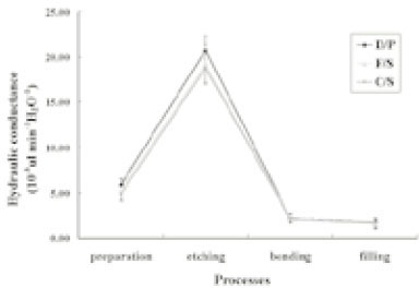
Fig. 3 The difference of fluid conductance according to the each process of compomer restoration.

Fig. 4 Degree of dye penetration. A,B. After etching. C,D. After applying the bonding agent(Prime&Bond NT). E,F. After compomer filling(F2000, Compoglass flow).
Cited by 1 articles
-
Effect of different air-drying time on the microleakage of single-step self-etch adhesives
Horieh Moosavi, Maryam Forghani, Esmatsadat Managhebi
Restor Dent Endod. 2013;38(2):73-78. doi: 10.5395/rde.2013.38.2.73.
Reference
-
1. McLean JW, Nicholson JW, Wilson AD. Proposed nomenclature for glass-ionomer dental cements and related materials. Quintessence Int. 1994. 25:587–589.2. Van Dijken JW. 3-year clinical evaluation of a compomer, a resin-modified glass ionomer and a resin composite in Class III restorations. Am J Dent. 1996. 9:195–198.3. Abdalla Al, Alhadainy HA, Garcia-Godoy F. Clinical evaluation of glass ionomers and compomers in Class V carious lesions. Am J Dent. 1997. 10:18–20.4. Kakabura A, Eliades G, Palahias G. Evaluation of the extent of the acid-base reaction on Dyract restorative material. J Dent Res. 1996. 75:1218. [Abstract 9].5. Gladys S, Van Meerbeek B, Braem M, Lambrechts P, Vangerle G. Comparative physico-mechanical characterization of new hybrid restorative materials with conventional glass-ionomer and resin composite restorative materials. J Dent Res. 1997. 76:883–894.
Article6. Triana R, Prado C, Garro J, Garcia-Godoy F. Dentin bond strength of fluoride-releasing materials. Am J Dent. 1994. 7:252–254.7. Ferrari M, Vichi A, Mannocci , Davidson CL. Sealing ability of two "compomers" applied with and without phosphoric acid treatment for Class V restorations in vivo. J Prosthet Dent. 1998. 79:131–135.
Article8. Tyas MJ. Clinical Evaluation of a polyacid-modified resin composite(compomer). Oper Dent. 1998. 23:77–80.9. Brackett WW, Gunnin TD, Gilpatrick RO, Browning WD. Microleakage of Class V compomer and light-curing glass ionomer restorations. J Prosthet Dent. 1998. 79:261–263.
Article10. Toledano M, Osorio E, Osorio R, Garcia-Godoy F. Microleakage of Class V resin-modified glass ionomer and compomer restorations. J Prosthet Dent. 1999. 81:610–615.
Article11. Attin T, Buchalla W, Kielbassa AM, Helwig E. Curing shrinkage and volumetric changes of rein-modified glass ionomer restorative materials. Dent Mater. 1995. 11:359–362.
Article12. Kidd EA. Microleakage: a review. J Dent. 1976. 4:199–206.13. Davidson CL. Resisting the curing contraction with adhesive composites. J Prosthet Dent. 1986. 55:446–447.
Article14. Feilzer AJ, de Gee AJ, Davidson CL. Relaxation of polymerization contraction shear stress by hygroscopic expansion. J Dent Res. 1990. 69:36–39.
Article15. Outhwaite WC, Livingston MJ, Pashley DH. Effects of changes in surface area, thickness, temperature and post-extraction time on human dentine permeability. Arch Oral Biol. 1976. 21:599–603.
Article16. Derkson GD, Pashley DH, Derkson ME. Microleakage measurement of selected restorative materials: A new in vitro method. J Prosthet Dent. 1986. 56:435–440.
Article17. Wu MK, Wesselink PR. Endodontic leakage studies reconsidered. part 1. Methodology, application and relevance. Int Endod J. 1993. 26:37–43.
Article18. Pommel L, Camps J. Effects of Pressure and Measurement time on the fluid filtration method in endodontics. J Endod. 2001. 27:256–258.
Article19. Otsuki M. Histological study on pulpal response to restorative composite resins and their ingredients. J Stomatol Soc Jpn. 1988. 55:203.
Article20. Ciucchi B, Bouillaquet S, Holz J, Pashley D. Dentinal fluid dynamics in human teeth, in vivo. J Endod. 1995. 21:191–194.
Article21. Kugel G, Perry RD, Hoang E, Ferrari M. Dyract compomer: Comparison of total etch vs. no etch technique. Gen Dent. 1998. 46:604–606.22. Ferrari M, Mannocci F, Kugel G, Garcia-Godoy F. Standardized microscopic evaluation of the bonding mechanism of NRC/Prime & Bond NT. Am J Dent. 1999. 12:77–83.23. Hansen SE, Swift EJ Jr, Krell KV. Permeability effects of two dentin adhesive systems. J Esthet Dent. 1992. 4:169–171.
Article24. Yoo HM, Park DS, Oh TS. Microleakage of class v compomer restorations. J Korean Acad Conserv Dent. 2000. 25:41–45.25. Jang HJ, Lee HJ, Hur B. Comparison of marginal leakage of wedge-shaped class v cavity according to restorative materials. J Korean Acad Conserv Dent. 2000. 25:56–62.26. Bourke AM, Walls AWG, McCabe JF. Light-activated glass polyalkenoate(ionomer) cements: The setting reaction. J Dent. 1992. 20:115–120.27. Darbyshire PA, Messer LB, Douglas WH. Microleakage in Class II composite restorations bonded to using thermal and load cycling. J Dent Res. 1988. 67:585–587.
Article28. Litkowski KJ, McDonald NJ, Swierczewski MA. A comparison of thermocycling methods for evaluating microleakage. J Dent Res. 1989. 68:Abstract 208.29. Hakimeh S, Vaidyanathan J. Microleakage of compomer Class V restorations: Effect of load cycling, thermal cycling, and cavity shape differences. J Prosthet Dent. 2000. 83:194–203.
Article
- Full Text Links
-
- Actions
-
Cited
- CITED
-
- Close
- Share
-
- Similar articles
-
- A study on the radiopacity of cavity lining materials for posterior composite resin restoration
- The Effects Of Desensitizing Agents And Tooth Brushing On Dentin Permeability, In Vitro
- The Effect of Hemostatic Solution on Dentin Permeability
- Effects on the tissue reaction using compomer & Ketac Silver in the maxillary furcation in the beagle dogs
- The effects of surface contamination by hemostatic agents on the shear bond strength of compomer
